. 2019 Sep 5;19(1):878.
doi: 10.1186/s12885-019-6087-1.

Inhibitory short peptides targeting EPS8/ABI1/SOS1 tri-complex suppress invasion and metastasis of ovarian cancer cells

Affiliations

Inhibitory short peptides targeting EPS8/ABI1/SOS1 tri-complex suppress invasion and metastasis of ovarian cancer cells

Xuechen Yu et al. BMC Cancer. .

Abstract

Background: We aimed to develop inhibitory short peptides that can prevent protein interactions of SOS1/EPS8/ABI1 tri-complex, a key component essential for ovarian cancer metastasis.

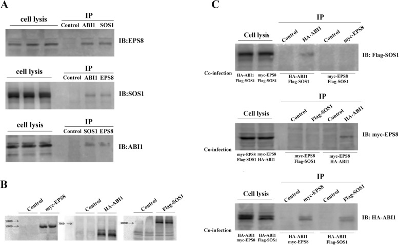
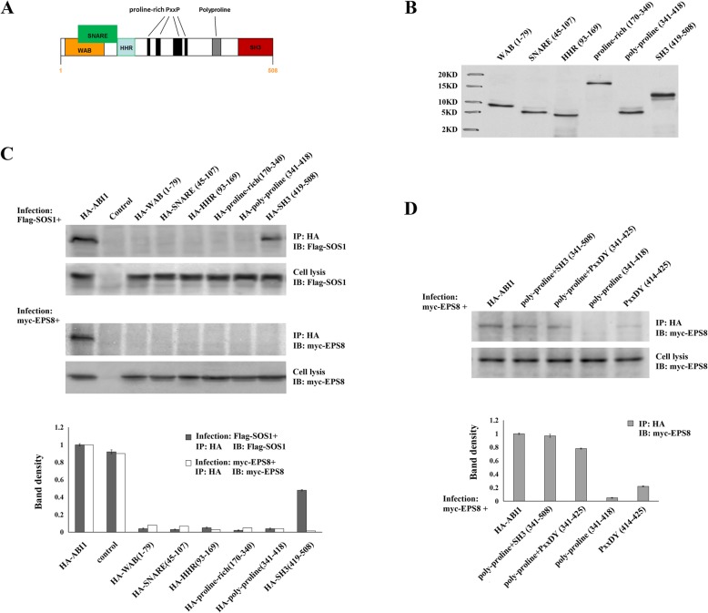
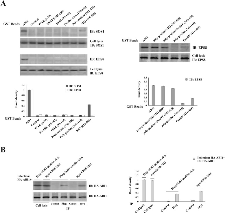
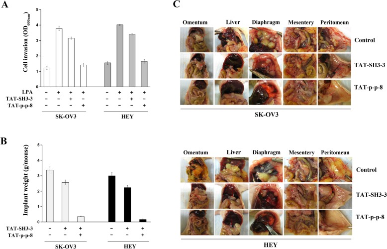
Methods: Plasmids containing various regions of HA-tagged ABI1 were co-transfected into ovarian cancer cells with Flag-tagged SOS1 or Myc-tagged EPS8. Co-immunoprecipitation and GST-pulldown assay were used to identify the regions of ABI1 responsible for SOS1 and EPS8 binding. Inhibitory short peptides of these binding regions were synthesized and modified with HIV-TAT sequence. The blocking effects of the peptides on ABI1-SOS1 or ABI1-EPS8 interactions in vitro and in vivo were determined by GST-pulldown assay. The capability of these short peptides in inhibiting invasion and metastasis of ovarian cancer cell was tested by Matrigel invasion assay and peritoneal metastatic colonization assay.

Results: The formation of endogenous SOS1/EPS8/ABI1 tri-complex was detected in the event of LPA-induced ovarian cancer cell invasion. In the tri-complex, ABI1 acted as a scaffold protein holding together SOS1 and EPS8. The SH3 and poly-proline+PxxDY regions of ABI1 were responsible for SOS1 and EPS8 binding, respectively. Inhibitory short peptides p + p-8 (ppppppppvdyedee) and SH3-3 (ekvvaiydytkdkddelsfmegaii) could block ABI1-SOS1 and ABI1-EPS8 interaction in vitro. TAT-p + p-8 peptide could disrupt ABI1-EPS8 interaction and suppress the invasion and metastasis of ovarian cancer cells in vivo.

Conclusions: TAT-p + p-8 peptide could efficiently disrupt the ABI1-EPS8 interaction, tri-complex formation, and block the invasion and metastasis of ovarian cancer cells.

Keywords: ABI1; EPS8; Inhibitory short peptide; Ovarian cancer metastasis; SOS1.


Conflict of interest statement

The authors declare that there are no conflicts of interest in this manuscript.

Figures

Fig. 1
Fig. 1
The formation of endogenous SOS1/EPS8/ABI1 tri-complex in ovarian cancer cell invasion. ABI1 served as a scaffold protein in SOS1/EPS8/ABI1 tri-complex. a. The metastatic ovarian cancer cell line SK-OV3 was treated with LPA for 5 min to induce cell invasion. Co-IP assay was employed to investigate the formation of endogenous SOS1/EPS8/ABI1 tri-complex after LPA stimulation. b. The Myc-tagged EPS8, HA-tagged ABI1 and Flag-tagged SOS1 recombinant plasmids were constructed. c. The OVCAR3 cells, which has no ABI1 expression, were infected by every two of the three expression plasmids (Myc-EPS8, HA-ABI1 and Flag-SOS1). Cell lysates were collected after LPA stimulation. The Co-IP assay was used to detect the interactions of ABI1-SOS1, ABI1-EPS8, and SOS1-EPS8
Fig. 2
Fig. 2
Characterizing regions in ABI1 mediating its interaction with SOS1 and EPS8. a. ABI1 was divided into the following regions: WAB (aa:1–79), SNARE (aa:54–108), HHR (aa:108–153), proline-rich (aa:153–331), poly-proline (aa:331–384) and SH3(aa:384–508). b. The plasmids containing HA-tagged various regions of ABI1 above were generated, using the pCDH-CMV-MCSEF1-Puro vector. The empty vector was used as control. c. The plasmids with different regions of ABI1 were co-infected into OVCAR3 cells with Flag-tagged SOS1 or Myc-tagged EPS8, respectively. Cell lysates were collected after LPA stimulation. Co-IP was performed to map out the regions in ABI1 responsible for ABI1-SOS1 or ABI1-EPS8 interactions. The results were quantified by ImageJ. The band density of each sample were normalized by cell lysis that detected the same protein and compared with control. d. We re-divided the C-terminal of ABI1 into the following regions: poly-proline+SH3 region (aa:331–508), poly-proline+PxxDY (aa:331–425)、poly-proline (aa:331–384)、PxxDY (aa:414–425). HA-tagged plasmids containing those regions were generated. Co-IP were performed to further identify which region mediated the ABI1-EPS8 interaction
Fig. 3
Fig. 3
Confirming the regions of ABI1 responsible for EPS8 or SOS1 binding. Characterizing regions in SOS1 and EPS8 mediating their interaction with ABI1. a. GST-pulldown assay was performed as follows. Recombinant GST-fused beads with various ABI1 fragments were incubated with LPA-treated OVCAR3 cell lysates and subsequently analyzed by immunoblotting to detect SOS1 or EPS8 expression. Results from above experiments also identified the SH3 and poly-proline+PxxDY as the regions in ABI1 responsible for SOS1 and EPS8 interaction, respectively. b. Flag-tagged proline-rich region of SOS1 (aa:1131–1333) and Myc-tagged SH3 region of EPS8 (aa:535–586) recombinant plasmids were generated, and then co-infected with HA-tagged ABI1 into OVCAR3 cells, respectively. Cell lysates were collected after LPA stimulation. The Co-IP assay was performed to determine the regions in SOS1 and EPS8 mediating their interactions with ABI1
Fig. 4
Fig. 4
Investigating inhibitory short peptides that can disrupt SOS1/EPS8/ABI1 tri-complex. Confirming the inhibitory capability of short peptides in vivo. a. we generated a series of synthetic inhibitory short peptides (each with 15 aa and overlapped by 5 aa) according to the poly-proline+PxxDY region of ABI1. After LPA stimulation, inhibitory short peptides were added to SK-OV3 cell lysates for 1 h prior to incubation with GST-ABI1 beads. GST-pulldown was employed to determine the effects of these peptides on ABI1-EPS81 binding. b. A series of synthetic inhibitory short peptides was generated (each is 25-amino acids long and overlapped by 5-amono acids) according to the SH3 region of ABI1. GST-pulldown was employed to determine the effects of these peptides on ABI1-SOS1 binding. c. The TAT-containing inhibitory short peptides or control peptides (10 μM) were added into the cell culture of SK-OV3 and incubate for different times. Then, GST-pulldown assay was performed to test the effects of TAT-containing inhibitory short peptides on ABI1-EPS8 interaction. d. The same experiments as above were performed to test the effects of TAT-containing inhibitory short peptides on ABI1-SOS1 interaction
Fig. 5
Fig. 5
Effects of inhibitory short peptides on the invasion and metastatic colonization of ovarian cancer cells. A. SK-OV3 and HEY cells were treated by TAT-fused peptides (TAT-p + p-8 or TAT-SH3–2) for 24 h before LPA-stimulation. Matrigel invasion assay was performed to test the suppressive capabilities of these inhibitory short peptides on cell invasion. Effect of inhibitory peptides on peritoneal metastatic colonization of ovarian cancer cells. B and C. SK-OV3 or HEY cells were intraperitoneally injected into nu/nu mice. Seventy-two hrs after injection, mice were intraperitoneally injected with PBS, TAT-fused scramble peptides or TAT-p + p-8 every 2 days for four weeks, respectively. By comparing the weight of metastatic implants from animals receiving inhibitory short peptides to those receiving PBS or scrambled control peptides, we estimated the capability of inhibitory short peptides in suppressing the metastasis of ovarian cancer cells

Similar articles

Cited by

References

    1. Siegel R, Ma J, Zou Z, Jemal A. Cancer statistics, 2014. CA Cancer J Clin. 2014;64(1):9–29. doi: 10.3322/caac.21208. - DOI - PubMed
    1. Ray-Coquard I, Morice P, Lorusso D, Prat J, Oaknin A, Pautier P, et al. ESMO Guidelines Committee. Non-epithelial ovarian cancer: ESMO Clinical Practice Guidelines for diagnosis, treatment and follow-up. Ann Oncol. 2018;29(Supplement_4):iv1–iv18. doi: 10.1093/annonc/mdy001. - DOI - PubMed
    1. Argento M, Hoffman P, Gauchez AS. Ovarian cancer detection and treatment: current situation and future prospects. Anticancer Res. 2008;28(5B):3135–3138. - PubMed
    1. Vaughan S, Coward JI, Bast RC, Jr, Berchuck A, Berek JS, Brenton JD, et al. Rethinking ovarian cancer: recommendations for improving outcomes. Nat Rev Cancer. 2011;11(10):719–725. doi: 10.1038/nrc3144. - DOI - PMC - PubMed
    1. Landen CN, Jr, Birrer MJ, Sood AK. Early events in the pathogenesis of epithelial ovarian cancer. J Clin Oncol. 2008;26(6):995–1005. doi: 10.1200/JCO.2006.07.9970. - DOI - PubMed