Case Reports
. 2019 Sep 5;19(1):199.
doi: 10.1186/s12886-019-1209-8.

Morphological characteristics of ocular toxoplasmosis and its regression pattern on swept-source optical coherence tomography angiography: a case report

Affiliations
Case Reports

Morphological characteristics of ocular toxoplasmosis and its regression pattern on swept-source optical coherence tomography angiography: a case report

Joong Hyun Park et al. BMC Ophthalmol. .

Abstract

Background: To report the successful treatment of ocular toxoplasmosis and present the use of multimodal imaging to describe the changes in ocular toxoplasmic lesions subsequent to treatment.

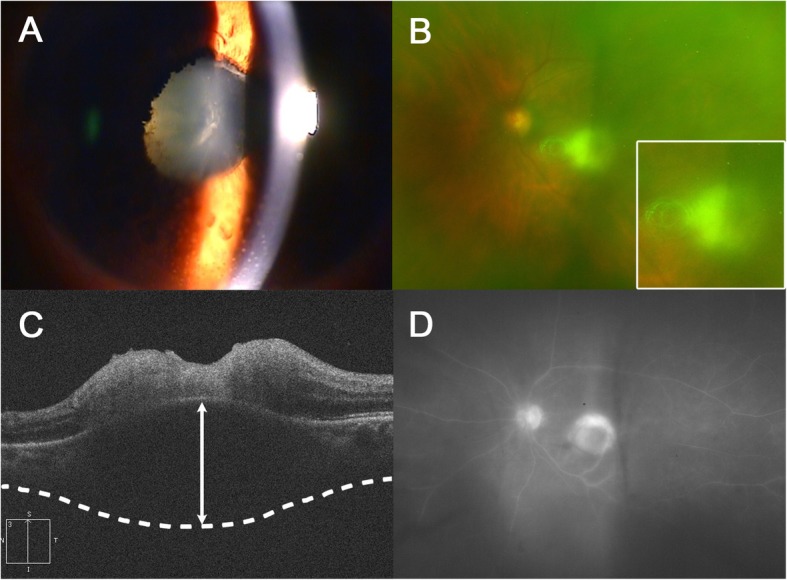
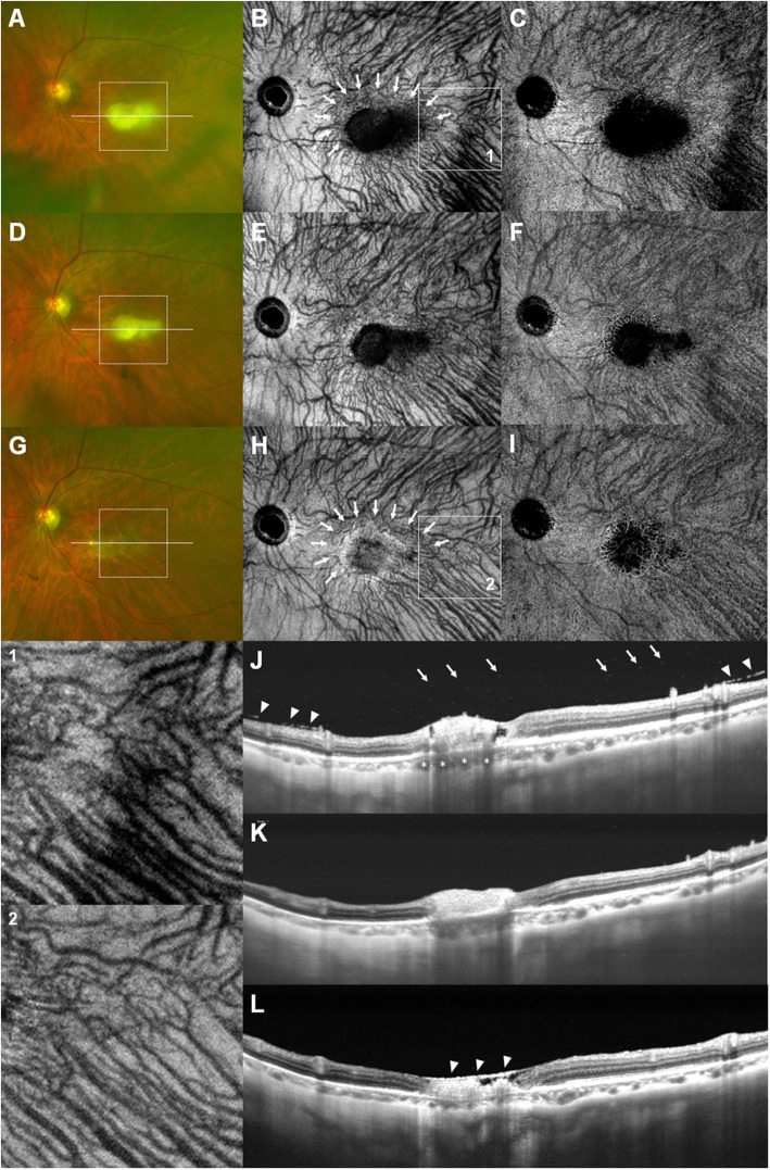
Case presentation: A 73-year-old female visited the clinic with decreased visual acuity in the left eye. Fundus examination showed severe vitreous haze with yellow-white infiltrates near the foveal center. Spectral-domain optical coherence tomography (SD-OCT) revealed disorganization of the retinal structure with markedly thickened choroid beneath the active lesion. Highly elevated serum titers of IgG antibodies against Toxoplasma gondii were observed. Topical and systemic steroids with oral Bactrim were administered after a diagnosis of ocular toxoplasmosis was made. After improvement in the severity of vitritis, structural en face swept-source optical coherence tomography (SS-OCT) imaging demonstrated diffuse choroidal dilation with many collateral vascular branches surrounding the active lesion. Eight intravitreal injections of clindamycin (1 mg/0.1 ml) were administered at 1- to 2-week intervals along with systemic antibiotics and steroids. After the treatment, the toxoplasmic lesion resolved to an atrophic chorioretinal scar. Dilated choroidal vessel size was normalized and collateral vascular branches were markedly constricted on structural en face SS-OCT images.

Conclusions: This is the first detailed report on the morphological changes in the choroidal vasculature surrounding ocular toxoplasmic lesions that were characterized using SS-OCT-A imaging. Multimodal imaging with SS-OCT-A can be valuable in clinical diagnosis as well as in clarifying the mechanism of choroidal structural changes in ocular toxoplasmosis.

Keywords: Case report; Morphological changes; Ocular toxoplasmosis; Swept-source optical coherence tomography angiography.


Conflict of interest statement

The authors declare that they have no competing interests.

Figures

Fig. 1
Fig. 1
Multi-modal imaging at initial presentation. a. Anterior segment photograph revealing mutton-fat keratic precipitates and peripupillary fibrous membrane with posterior synechiae. b. Wide-field fundus photography demonstrating macular yellow-white infiltrates with vitreous haze (white frame, magnified image). c. Disorganization of the retinal structure with markedly thickened choroid (white double-headed arrow) as observed on SD-OCT. d. Wide-field fluorescein angiography showing optic disc leakage and hyperfluorescence of the active lesion
Fig. 2
Fig. 2
Fundus photographs (a, d, g), structural en face SS-OCT images (b, e, h), SS-OCT-A images (c, f, i), and SS-OCT B-scans (j, k, l) during the course of treatment. At 4 weeks after treatment, a. Vitreous haze decreased, but yellow-white infiltrate remained near the foveal center. b. Many collateral vascular branches surrounding the lesion (white arrows) and diffuse choroidal dilation, which is more remarkable in magnified image (Box 1) are shown. c. SS-OCT-A image showed congested choroidal vasculature. d, e, f. After two intravitreal injections of clindamycin, the size of macular infiltrates decreased with more discrete margins. g. After 3.5 months of additional treatment, the macular lesion changed to chorioretinal scar. h, i. Normalization of dilated choroidal vessels and constriction of collateral branch vessels around the lesion (white arrows), which is more remarkable in magnified image (Box 2) are seen. j. An SS-OCT B-scan through the lesion (white line on the fundus photograph) demonstrated disruption of the neurosensory retina, interruption of the photoreceptor inner and outer segment junction and RPE elevation. Multiple hyperreflective dots in the vitreous cavity indicating severe vitritis (white arrows), thickened posterior hyaloid (white arrowheads), and dilated Haller’s layer vessels (white asterisks) were also noted. k. The choroidal thickness decreased further. l. Atrophic retinal thinning, overlying thickened posterior hyaloid (white arrowheads), and pronounced choroidal thinning are seen

Similar articles

Cited by

References

    1. Holland Gary N. Ocular toxoplasmosis: a global reassessment. American Journal of Ophthalmology. 2003;136(6):973–988. doi: 10.1016/j.ajo.2003.09.040. - DOI - PubMed
    1. Rothova A. Ocular manifestations of toxoplasmosis. Curr Opin Ophthalmol. 2003;14:384–388. doi: 10.1097/00055735-200312000-00011. - DOI - PubMed
    1. Bosch-Driessen LE, Berendschot TT, Ongkosuwito JV, Rothova A. Ocular toxoplasmosis: clinical features and prognosis of 154 patients. Ophthalmology. 2002;109:869–878. doi: 10.1016/S0161-6420(02)00990-9. - DOI - PubMed
    1. Jones JL, Kruszon-Moran D, Wilson M, McQuillan G, Navin T, McAuley JB. Toxoplasma gondii infection in the United States: seroprevalence and risk factors. Am J Epidemiol. 2001;154:357–365. doi: 10.1093/aje/154.4.357. - DOI - PubMed
    1. Chen KC, Jung JJ, Engelbert M. Single acquisition of the vitreous, retina and choroid with swept-source optical coherence tomography in acute toxoplasmosis. Retin Cases Brief Rep. 2016;10:217–220. doi: 10.1097/ICB.0000000000000230. - DOI - PubMed

Publication types

MeSH terms