Review
. 2019 Sep 5;19(1):274.
doi: 10.1186/s12888-019-2247-8.

Case report: a giant arachnoid cyst masking Alzheimer's disease

Affiliations
Review

Case report: a giant arachnoid cyst masking Alzheimer's disease

Anna-Sophia Wahl et al. BMC Psychiatry. .

Abstract

Background: Intracranial arachnoid cysts are usually benign congenital findings of neuroimaging modalities, sometimes however, leading to focal neurological and psychiatric comorbidities. Whether primarily clinically silent cysts may become causally involved in cognitive decline in old age is neither well examined nor understood.

Case presentation: A 66-year old caucasian man presenting with a giant left-hemispheric frontotemporal cyst without progression of size, presented with slowly progressive cognitive decline. Neuropsychological assessment revealed an amnestic mild cognitive impairment (MCI) without further neurological or psychiatric symptoms. The patient showed mild medio-temporal lobe atrophy on structural MRI. Diffusion tensor and functional magnetic resonance imaging depicted a rather sustained function of the strongly suppressed left hemisphere. Amyloid-PET imaging was positive for increased amyloid burden and he was homozygous for the APOEε3-gene. A diagnosis of MCI due to Alzheimer's disease was given and a co-morbidity with a silent arachnoid cyst was assumed. To investigate, if a potentially reduced CSF flow due to the giant arachnoid cyst contributed to the early manifestation of AD, we reviewed 15 case series of subjects with frontotemporal arachnoid cysts and cognitive decline. However, no increased manifestation of neurodegenerative disorders was reported.

Conclusions: With this case report, we illustrate the necessity of a systematic work-up for neurodegenerative disorders in patients with arachnoid cysts and emerging cognitive decline. We finally propose a modus operandi for the stratification and management of patients with arachnoid cysts potentially susceptive for cognitive dysfunction.

Keywords: Alzheimer’s disease; Arachnoid cysts; Cognitive decline; Functional neuro-imaging; Neural plasticity.


Conflict of interest statement

The authors have no patents pending or financial conflicts to disclose. This research did not receive any specific grant from funding agencies in the public, commercial, or not-for-profit sectors.

Figures

Fig. 1
Fig. 1
Brain MRI showing a large left frontotemporal cerebrospinal fluid-filled cyst with a cross-sectional area of 6 × 14 cm compatible with a Type III arachnoid cyst after the Galassi classification of middle cranial fossa arachnoid cysts: a FLAIR axial, b T2 TSE sagittal, and c T1 SE coronal view
Fig. 2
Fig. 2
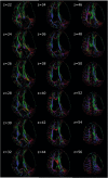
Images of first eigenvector overlaid on fractional anisotropy (FA) images. The brightness of the vector colors in each voxel is modulated according to the values in the corresponding FA image, indicating the axis of diffusion. The colors red, green and blue correspond to the directions left/right, anterior/posterior and inferior/superior, respectively. The slice position in axial direction is given by its z coordinate next to the image
Fig. 3
Fig. 3
Functional magnetic resonance imaging results overlaid on the inflated left and right hemisphere reconstruction. The rows 1 to 5 depict activation patterns in response to digit 1 (first row) to digit 5 (last row). The first and second columns depict activation in the left (first column) and right hemisphere (second column) in response to left hand stimulation. The third and fourth columns depict activation in the left (third column) and right hemisphere (fourth column) in response to right hand stimulation. Activation is shown as ‘significances’, ie, −log10(p). So, for p = .001, sig = 3. Colour code ranges from 3 to 10 as illustrated by the color bar in the center. Additionally outlines of pre- and postcentral gyrus are depicted in blue and yellow, corresponding to parcellation results depicted in Additional file 1: Figure S1
Fig. 4
Fig. 4
Coronal and horizontal brain slices of Amyloid-PET imaging. F18-FBB (Florbetaben) tracer was used depicting amyloid-beta burden in both grey and white matter. The border between grey and white matter is indistinguishable thus indicating a pathological cortical amyloid-beta deposit

Similar articles

Cited by

References

    1. Weber F, Knopf H. Incidental findings in magnetic resonance imaging of the brains of healthy young men. J Neurol Sci. 2006;240(1–2):81–84. doi: 10.1016/j.jns.2005.09.008. - DOI - PubMed
    1. Zwagerman NT, Pardini J, Mousavi SH, Friedlander RM. A refractory arachnoid cyst presenting with tremor, expressive dysphasia, and cognitive decline. Surg Neurol Int. 2016;7(Suppl 15):S431–S433. - PMC - PubMed
    1. Al-Holou WN, Yew AY, Boomsaad ZE, Garton HJ, Muraszko KM, Maher CO. Prevalence and natural history of arachnoid cysts in children. J Neurosurg Pediatr. 2010;5(6):578–585. doi: 10.3171/2010.2.PEDS09464. - DOI - PubMed
    1. Katzman GL, Dagher AP, Patronas NJ. Incidental findings on brain magnetic resonance imaging from 1000 asymptomatic volunteers. JAMA : the journal of the American Medical Association. 1999;282(1):36–39. doi: 10.1001/jama.282.1.36. - DOI - PubMed
    1. Vernooij MW, Ikram MA, Tanghe HL, Vincent AJ, Hofman A, Krestin GP, et al. Incidental findings on brain MRI in the general population. N Engl J Med. 2007;357(18):1821–1828. doi: 10.1056/NEJMoa070972. - DOI - PubMed