. 2019 Sep 5;19(1):64.
doi: 10.1186/s12896-019-0554-2.

Intracellular human antibody fragments recognizing the VP35 protein of Zaire Ebola filovirus inhibit the protein activity

Affiliations

Intracellular human antibody fragments recognizing the VP35 protein of Zaire Ebola filovirus inhibit the protein activity

Michela Flego et al. BMC Biotechnol. .

Abstract

Background: Ebola hemorrhagic fever is caused by the Ebola filovirus (EBOV), which is one of the most aggressive infectious agents known worldwide. The EBOV pathogenesis starts with uncontrolled viral replication and subversion of both the innate and adaptive host immune response. The multifunctional viral VP35 protein is involved in this process by exerting an antagonistic action against the early antiviral alpha/beta interferon (IFN-α/β) response, and represents a suitable target for the development of strategies to control EBOV infection. Phage display technology permits to select antibodies as single chain Fragment variable (scFv) from an artificial immune system, due to their ability to specifically recognize the antigen of interest. ScFv is ideal for genetic manipulation and to obtain antibody constructs useful for targeting either antigens expressed on cell surface or intracellular antigens if the scFv is expressed as intracellular antibody (intrabody) or delivered into the cells.

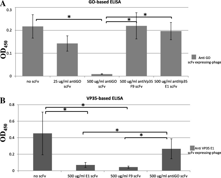
Results: Monoclonal antibodies (mAb) in scFv format specific for the EBOV VP35 were isolated from the ETH-2 library of human recombinant antibodies by phage display technology. Five different clones were identified by sequencing, produced in E.coli and expressed in CHO mammalian cells to be characterized in vitro. All the selected scFvs were able to react with recombinant VP35 protein in ELISA, one of the scFvs being also able to react in Western Blot assay (WB). In addition, all scFvs were expressed in cell cytoplasm as intrabodies; a luciferase reporter gene inhibition assay performed in A549 cells showed that two of the scFvs can significantly hamper the inhibition of the IFN-β-induced RIG-I signaling cascade mediated by EBOV VP35.

Conclusion: Five antibodies in scFv format recognize an active form of EBOV VP35 in ELISA, while one antibody also recognizes VP35 in WB. Two of these scFvs were also able to interfere with the intracellular activity of VP35 in a cell system in vitro. These findings suggest that such antibodies in scFv format might be employed to develop therapeutic molecules able to hamper EBOV infections.

Keywords: Intrabody; VP35; Zaire ebolavirus; scFv.


Conflict of interest statement

The authors declare that they have no competing interests.

Figures

Fig. 1
Fig. 1
Reactivity of the scFvs against the recombinant EBOV VP35 in ELISA. The IPTG-induced bacterial supernatants of individual colonies from the third round of selection were tested in 96-well microtiter plates coated with the recombinant VP35 protein as an antigen. The cut-off value separating positive from negative samples was calculated as 3 standard deviation values (SD) above the mean, of the values obtained using the irrelevant anti-GO scFv (OD450 = 0.079). The five different positive clones isolated, identified by sequence analysis, are indicated
Fig. 2
Fig. 2
Molecular characterization of the CDR3 belonging to the selected anti-EBOV VP35 scFvs. a Schematic representation of the scFv gene in the phage display cassette showing the position of the variable CDR3 in the VH and VL chains. Lac p: Lac promoter; PelB: peptide leader for secretion in the bacterial periplasm. b Sequences of the DP47 VH gene and DPL16 VL gene are shown. CDR1, CDR2, CDR3, identified through IMGT (international ImMunoGeneTics information system)/V-Quest analysis are underlined. c Amino acid composition of the CDR3 regions in the VH and VL domains of the five most reactive anti-VP35 scFvs
Fig. 3
Fig. 3
a Reactivity of the anti-VP35 scFv clones in ELISA. Either the recombinant VP35 (blue bars) or the recombinant glucose oxidase (GO, red bars) were used as antigens. The anti-GO scFv was used as control (ctrl) for detection in parallel with the anti-VP35 scFvs. The mean values and the SD of a representative experiment out of at least three, performed in triplicate, are reported. b Reactivity of the anti-VP35 scFv clones in immunoblot assay. Recombinant VP35 protein and GO antigen as a control were analyzed by SDS-PAGE, blotted onto nitrocellulose membrane and cut into strips. Each strip was incubated with the supernatant deriving from a bacterial clone producing one of the anti-VP35 scFvs as indicated, and the antigen-antibody reaction revealed by ECL. The GO antigen detection by ctrl scFv and the recombinant VP35 detection by anti-His mAb, were used as positive controls. The molecular weights (MW) of the bands corresponding to GO and VP35 protein are indicated by arrows
Fig. 4
Fig. 4
a Expression of the monoclonal scFv in eukaryotic cells. WB analysis showing the transient expression of the A10, H7, B10, F9 and E1 scFvs in CHO cells 72 h after transfection with the respective recombinant pTarget plasmids. Specific bands corresponding to scFvs (about 27 kDa) are detected for all clones; other bands of higher molecular mass are present in all the lanes and are a cross-reactivity as evidenced by their presence also in CHO control lane. Molecular mass of the marker is indicated. b Controls used in the luciferase reporter gene assay. The histogram shows the effect of transfection procedures on the IFN-β promoter expression. Twenty-four hours after co-transfection with pGL IFN-β luc and pcDNA3 or pcDNA3 EBOV wtVP35 expression vectors, cells were transfected with the ctrl anti-GO scFv pTarget. The next day, cells were additionally transfected with IAV vRNA. The results from four independent experiments performed in triplicate are shown as fold induction of stimulated samples with respect to unstimulated control. c. ScFv against EBOV VP35 protein effect in the luciferase reporter gene assay. Twenty-four hours after co-transfection with pGL IFN-β luc and pcDNA3 or pcDNA3 EBOV wtVP35 expression vector, cells were additionally transfected with different scFv p Target vectors and next day additionally transfected with IAV vRNA. The results from three independent experiments are shown as a percentage of the IFNβ promoter induction. Bars indicate the mean ± SD; asterisks indicate a significant difference: **P < 0.01 and ***P < 0.005 (two-tailed unpaired Student’s t-test, n = 3)
Fig. 5
Fig. 5
Competitive ELISA for characterization of the scFv binding to EBOV VP35. The binding of the scFv-expressing phages (5 × 109 tu/ml) was measured, both in the absence and in the presence of the competitor soluble non-phage-fused scFvs at the maximum concentration of 500 μg/ml. An anti -M13 PVIII coat protein mAb conjugated to HRP was used for phage detection. On the x-axis the concentrations of scFvs used for the competition assay are reported, on the y-axis, the OD450 phage ELISA values are reported. a Competitive ELISA using the anti-GO scFv-expressing phages. The binding of anti-GO scFv-expressing phages to plate coated with GO protein is shown: in the absence of competitor soluble non-phage-fused scFvs; in the presence of their own anti-GO soluble non-phage-fused scFvs at the concentration of 500 μg/ml or 25 μg/ml; in the presence of the anti-VP35 F9, and E1 soluble non-phage-fused scFvs, at the concentration of 500 μg/ml. The higher concentration of anti-GO soluble non-phage-fused scFvs shows to compete with itself. The mean values and the SD of an experiment performed in triplicate are reported. Binding in the presence of soluble non-phage-fused scFvs was compared to binding in presence of the control soluble non-phage-fused scFvs or in their absence using Student’s t-test. *p < 0.05; b Competitive ELISA using anti-EBOV VP35 scFv E1 expressing-phages. The binding of E1 anti-VP35 scFv-expressing phages to plate coated with VP35 protein is shown: in the absence of competitor soluble non-phage-fused scFvs; in the presence of their own E1 soluble non-phage-fused scFvs at the concentration of 500 μg /ml; in the presence of F9 soluble non-phage-fused scFvs at the concentration of 500 μg/ml; in the presence of the control anti-GO soluble non-phage-fused scFvs at the concentration of 500 μg/ml. The mean values and the SD of an experiment performed in triplicate are reported. Binding in the presence of soluble non-phage-fused scFvs was compared to binding in the presence of the control soluble non-phage-fused scFvs or in their absence using Student’s t-test. *p < 0.05

References

    1. Feldmann H, Geisbert TW. Ebola haemorrhagic fever. Lancet. 2011;377:849–862. doi: 10.1016/S0140-6736(10)60667-8. - DOI - PMC - PubMed
    1. Malvy D, McElroy AK, de Clerk H, Gunther S, van Griensven J. Ebola virus disease. Lancet. 2019;393:936–948. doi: 10.1016/S0140-6736(18)33132-5. - DOI - PubMed
    1. Goldstein T, Anthony SJ, Gbakima A, Bird BH, Bangura J, Tremeau-Bravard A, Belaganahalli MN, Wells HL, Dhanota JK, Liang E, Grodus M, Jangra RH, Dejesus VA, Lasso G, Smith BR, Jambai A, Kamara BO, Kamara S, Bangura W, Monagin C, Shapira S, Johnson CK, Saylors K, Rubin EM, Chandran K, Lipkin WI, Mazet J. The discovery of a new Ebolavirus, Bombali virus, adds further support for bats as hosts of Ebolaviruses. Int J Infect Dis. 2019;79(Supp 1):4–5. doi: 10.1016/j.ijid.2018.11.030. - DOI - PubMed
    1. Geisbert TW, Hensley LE. Ebola virus: new insights into disease aetiopathology and possible therapeutic interventions. Expert Rev Mol Med. 2004;6:1–24. doi: 10.1017/S1462399404008300. - DOI - PubMed
    1. Mohamadzadeh M, Chen L, Schmaljohn AL. How Ebola and Marburg viruses battle the immune system. Nat Rev Immunol. 2007;7:556–567. doi: 10.1038/nri2098. - DOI - PubMed

Publication types

MeSH terms