. 2019 Nov 5;9(11):3623-3630.
doi: 10.1534/g3.119.400633.

Regulation of Drosophila Intestinal Stem Cell Proliferation by Enterocyte Mitochondrial Pyruvate Metabolism

Affiliations

Regulation of Drosophila Intestinal Stem Cell Proliferation by Enterocyte Mitochondrial Pyruvate Metabolism

Dona R Wisidagama et al. G3 (Bethesda). .

Abstract

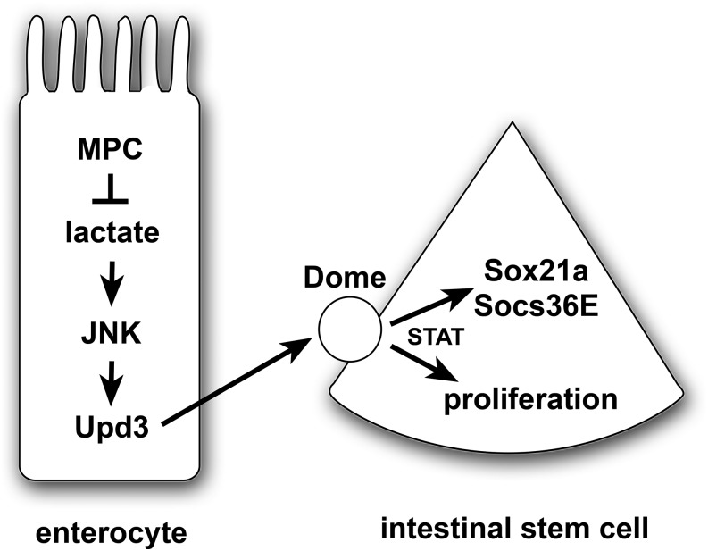
Multiple signaling pathways in the adult Drosophila enterocyte sense cellular damage or stress and signal to intestinal stem cells (ISCs) to undergo proliferation and differentiation, thereby maintaining intestinal homeostasis. Here we show that misregulation of mitochondrial pyruvate metabolism in enterocytes can stimulate ISC proliferation and differentiation. Our studies focus on the Mitochondrial Pyruvate Carrier (MPC), which is an evolutionarily-conserved protein complex that resides in the inner mitochondrial membrane and transports cytoplasmic pyruvate into the mitochondrial matrix. Loss of MPC function in enterocytes induces Unpaired cytokine expression, which activates the JAK/STAT pathway in ISCs, promoting their proliferation. Upd3 and JNK signaling are required in enterocytes for ISC proliferation, indicating that this reflects a canonical non-cell autonomous damage response. Disruption of lactate dehydrogenase in enterocytes has no effect on ISC proliferation but it suppresses the proliferative response to a loss of enterocyte MPC function, suggesting that lactate contributes to this pathway. These studies define an important role for cellular pyruvate metabolism in differentiated enterocytes to maintain stem cell proliferation rates.

Keywords: intestinal homeostasis; metabolism; mitochondria; stem cells.


Figures

Figure 1
Figure 1
Reduced mitochondrial pyruvate metabolism in enterocytes increases stem cell proliferation in a non-autonomous manner. (A) The EC-specific myo1A-GAL4 driver was used to target RNAi for mCherry (control), dMPC1, or PDH-E1 in the presence of Tub-GAL80ts. The number of cells staining positive for phosphorylated histone H3 (pHH3+ cells) was quantified per intestine after shifting 4-5 day old animals to 29°C for seven days. Data are plotted as the mean ± SEM, n ≥ 20 animals for each condition, ****P ≤ 0.0001. (B) Genes that encode key components in the JAK/STAT signaling pathway increase their expression in myo1A > dMPC1-RNAi intestines. Data from Supplementary Table 1 is depicted for genes involved in JAK/STAT signaling in the intestine, showing their rank order in the RNA-seq dataset and fold-change increase in expression relative to the control.
Figure 2
Figure 2
MPC loss of function in enterocytes requires UPD3 and JNK to indirectly control stem cell proliferation. The myo1A-GAL4 driver was used to target RNAi for mCherry (control) (A,C) or dMPC1 (B,D) in the presence of Tub-GAL80ts. The upd3.1-lacZ reporter (gray, A-D) and 10xSTAT92E-DGFP reporter (green, C,D) were used to detect Upd3 expression and JAK/STAT activation, respectively, by immunohistochemistry. Proliferating ISCs are marked by staining for pHH3+ cells (red) and nuclei are stained with DAPI (blue). Panels 2A,B and 2C,D are from independent experiments and depict the R4 region of the intestine. Scale bars represent 50 µm (A,B) and 20 µm (C,D). (E) The myo1A-GAL4 driver was used to target RNAi for mCherry (control), dMPC1, PDH-E1, or upd3, either alone or in combination as shown, in the presence of Tub-GAL80ts. The number of cells staining positive for phosphorylated histone H3 (pHH3+ cells) was quantified per intestine after shifting 4-5 day old animals to 29°C for seven days. Data are plotted as the mean ± SEM, n ≥ 20 animals for each condition. (F) The myo1A-GAL4 driver was used to target mCherry RNAi (control), dMPC1 RNAi, PDH-E2 RNAi, or BskDN expression, either alone or in combination as shown, in the presence of Tub-GAL80ts. The number of cells staining positive for phosphorylated histone H3 (pHH3+ cells) was quantified per intestine after shifting 4-5 day old animals to 29°C for seven days. Data are plotted as the mean ± SEM, n ≥ 20 animals for each condition. ****P ≤ 0.001, *P ≤ 0.05.
Figure 3
Figure 3
dMPC1 mutants have increased glycolytic intermediates and reduced TCA cycle intermediates. GC/MS metabolomic profiling of controls (blue bars) and dMPC1 mutants (red bars) on a diet containing 9% sugar and 8% yeast. Data are graphically represented as a box plot, with the box representing the lower and upper quartiles, the horizontal line representing the median, and the bars representing the minimum and maximum data points. Mutant values are normalized to the control and fold changes in metabolite levels are displayed. n = 4 independent experimental collections. ****P ≤ 0.0001, ***P ≤ 0.001, **P ≤ 0.01 *P ≤ 0.05.
Figure 4
Figure 4
LDH is required in enterocytes for the non-autonomous effect of MPC loss on stem cell proliferation. (A-F) The myo1A-GAL4 driver was used to target RNAi for mCherry (A), dMPC1 (B,E), PDH-E1 (C,F), and LDH (D-F) in the presence of Tub-GAL80ts. The upd3.1-lacZ reporter (gray) was used to detect Upd3 expression, proliferating ISCs are marked by staining for pHH3+ cells (red), and nuclei are stained with DAPI (blue). Images depict the R4 region of the intestine. Scale bar represents 50 µm. (G) The myo1A-GAL4 driver was used to target RNAi for mCherry (control), LDH, dMPC1, or PDH-E1 in the presence of Tub-GAL80ts. The number of cells staining positive for phosphorylated histone H3 (pHH3+ cells) was quantified per intestine after shifting 4-5 day old animals to 29°C for seven days. Data are plotted as the mean ± SEM, n ≥ 20 animals for each condition. ****P ≤ 0.0001.
Figure 5
Figure 5
A model for the non-autonomous regulation of ISC proliferation by enterocyte pyruvate metabolism. A loss of the MPC in enterocytes results in increased lactate production by LDH, which activates the JNK stress signaling pathway and Upd3 expression. This secreted cytokine acts through the Dome receptor in ISCs to promote JAK/STAT signaling, resulting in up-regulation of Sox21a and Socs36E as well as stem cell proliferation. Note that this is a speculative model that is intended to summarize the conclusions from this study in the context of the well-established JNK/STAT stress response pathway in the intestine.

References

    1. Bach E. A., Ekas L. A., Ayala-Camargo A., Flaherty M. S., Lee H. et al. , 2007. GFP reporters detect the activation of the Drosophila JAK/STAT pathway in vivo. Gene Expr. Patterns 7: 323–331. 10.1016/j.modgep.2006.08.003 - DOI - PubMed
    1. Beebe K., Lee W. C., and Micchelli C. A., 2010. JAK/STAT signaling coordinates stem cell proliferation and multilineage differentiation in the Drosophila intestinal stem cell lineage. Dev. Biol. 338: 28–37. 10.1016/j.ydbio.2009.10.045 - DOI - PubMed
    1. Bonfini A., Liu X., and Buchon N., 2016. From pathogens to microbiota: How Drosophila intestinal stem cells react to gut microbes. Dev. Comp. Immunol. 64: 22–38. 10.1016/j.dci.2016.02.008 - DOI - PubMed
    1. Bricker D. K., Taylor E. B., Schell J. C., Orsak T., Boutron A. et al. , 2012. A mitochondrial pyruvate carrier required for pyruvate uptake in yeast, Drosophila, and humans. Science 337: 96–100. 10.1126/science.1218099 - DOI - PMC - PubMed
    1. Buchon N., Broderick N. A., Chakrabarti S., and Lemaitre B., 2009a Invasive and indigenous microbiota impact intestinal stem cell activity through multiple pathways in Drosophila. Genes Dev. 23: 2333–2344. 10.1101/gad.1827009 - DOI - PMC - PubMed

Publication types