. 2019 Oct 1;33(19-20):1361-1366.
doi: 10.1101/gad.326314.119. Epub 2019 Sep 5.

USP21 deubiquitinase promotes pancreas cancer cell stemness via Wnt pathway activation

Affiliations

USP21 deubiquitinase promotes pancreas cancer cell stemness via Wnt pathway activation

Pingping Hou et al. Genes Dev. .

Abstract

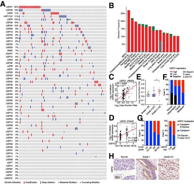
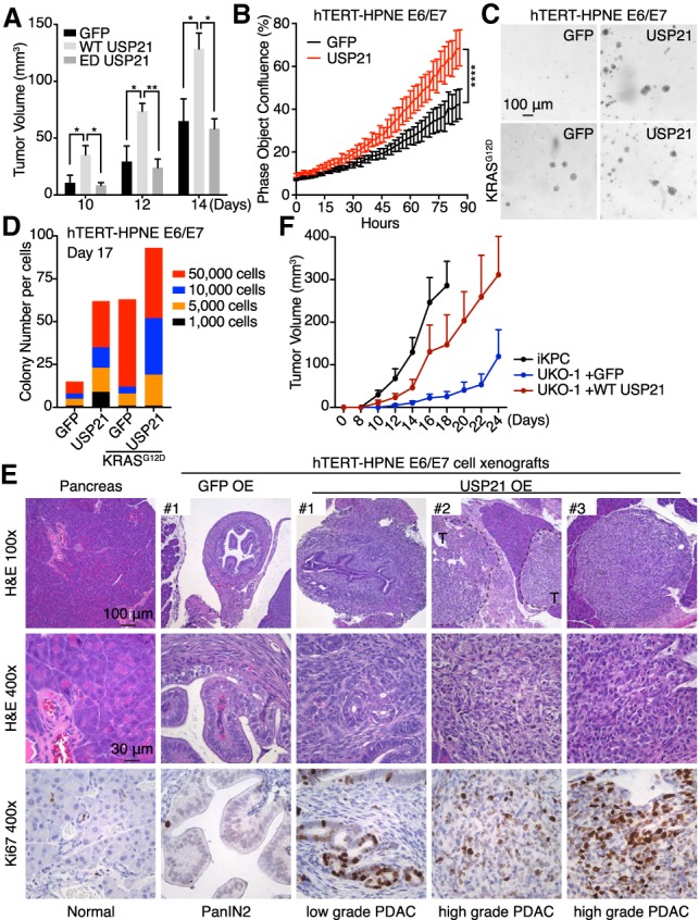
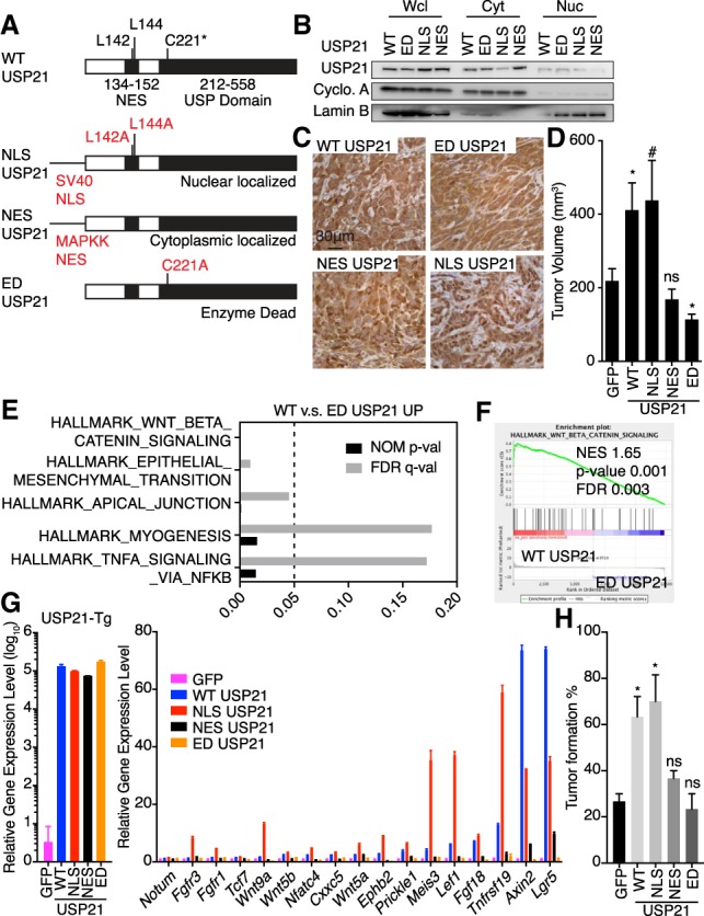
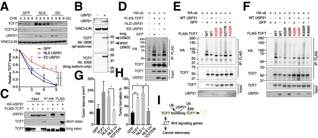
The ubiquitin-specific protease (USP) family is the largest group of cysteine proteases. Cancer genomic analysis identified frequent amplification of USP21 (22%) in human pancreatic ductal adenocarcinoma (PDAC). USP21 overexpression correlates with human PDAC progression, and enforced expression of USP21 accelerates murine PDAC tumor growth and drives PanIN to PDAC progression in immortalized human pancreatic ductal cells. Conversely, depletion of USP21 impairs PDAC tumor growth. Mechanistically, USP21 deubiquitinates and stabilizes the TCF/LEF transcription factor TCF7, which promotes cancer cell stemness. Our work identifies and validates USP21 as a PDAC oncogene, providing a potential druggable target for this intractable disease.

Keywords: TCF7; USP21; Wnt pathway; cancer stemness; deubiquitinase; pancreatic cancer.


Figures

Figure 1.
Figure 1.
USP21 is up-regulated in PDAC. (A,B) Genetic alternations of USPs in UTSW PDAC data set (A) and pan-cancer analysis (B). (C,D) USP21 mRNA level positively correlates with copy number (C) and GISTIC scores (D) in TCGA PAAD data set. (E) USP21 copy number positively correlates with PDAC tumor grade analyzed in Oncomine. (F) USP21 expression positively correlates with PDAC progression by TMA analysis. (G) USP21 nuclear localization positively correlates with tumor grade and USP21 expression by TMA analysis. (H) Representative IHC images of USP21 in TMA tissues.
Figure 2.
Figure 2.
USP21 promotes pancreatic tumor growth. (A) Tumor growth comparison of GFP, WT-, and ED-USP21 OE iKPC cells (n = 5). Two-hundred-thousand cells were injected subcutaneously in nude mice. Tumor sizes were measured on days 10, 12, and 14. (B) Cell proliferation comparison of GFP-HPNE and USP21-HPNE cells (n = 6). (C,D) Representative images (C) and colony quantification (D) of anchorage-independent colony formation of hTERT-HPNE E6/E7 and hTERT-HPNE E6/E7-KRASG12D cells overexpressing WT-USP21 and GFP. (E) Characterization of xenograft masses from orthotopically injected GFP-HPNE and USP21-HPNE cells in NSG mice (n = 4). Proliferative cells were positively stained by Ki67. (T) Tumor. (F) Knockout of Usp21 impaired iKPC tumor growth (n = 5). For A, B, and F, data are represented as mean ± SEM.
Figure 3.
Figure 3.
Nuclear function of USP21 activates Wnt pathway and increases cancer cell stemness. (A) Plasmid construction for specific subcellular localization of USP21. (B) Validation of subcellular localization of USP21 variants. (C) Representative IHC images of USP21 localization in iKPC cells overexpressing USP21 variants. (D) Comparison of tumor growth between iKPC cells overexpressing GFP and USP21 variants (n = 5). (E) GSEA of pathways enriched in WT-USP21 versus ED-USP21 OE iKPC cells. (F) Enrichment of Wnt pathway in WT-USP21 versus ED-USP21 OE iKPC cells. (G) RT-PCR validation of Wnt pathway genes comparing iKPC cells overexpressing GFP and USP21 variants (n = 3). Data are represented as mean ± SD. (H) Limited dilution assay comparing iKPC cells overexpressing GFP and USP21 variants. n = 3 independent experiments. For D and H, data are represented as mean ± SEM. Comparison is between GFP control with other groups as indicated.
Figure 4.
Figure 4.
TCF7 mediates tumor-promoting function of USP21. (A) CHX protein chasing assay of TCF7 and TCF7L2 in GFP, NLS-USP21, and ED-USP21 OE iKPC cells. Data are represented as mean ± SD (n = 3). (B) Western blot analysis of TCF7 isoforms in iKPC cells. (C) Co-IP analysis between WT-USP21 and TCF7 in 293T cells. Arrowhead indicates the correct band size. (D) Ubiquitination assay of TCF7 in GFP, NLS- USP21, and ED-USP21 OE 293T cells. (E) Ubiquitination assay of TCF7 in 293T cells overexpressing WT TCF7 and TCF7 mutants. (F) Ubiquitination assay in 293T cells overexpressing WT TCF7 and TCF7 mutants with and without USP21 cotransfection. (G) Tumor growth comparison of iKPC cells overexpressing indicated ORFs (n = 4). (H) Limited dilution assay comparing iKPC cells overexpressing indicated ORFs. n = 3 independent experiments. (I) Schematic graph of the USP21 regulation of TCF7 to promote cancer stemness in PDAC. Statistical analysis was performed between indicated groups. For G and H, data are represented as mean ± SEM.
Figure 5.
Figure 5.
USP21 stabilizes TCF7 and promotes tumor growth via TCF7 in human PDAC cells. (A) Comparison of TCF7 protein expression in GFP-HPNE and USP21-HPNE xenografts by IHC. (B) CHX assay of TCF7 in GFP and USP21 OE Mia PaCa-2 cells (n = 2). Data are represented as mean ± SD. (C) Tumor growth comparison of MIA PaCa-2 cells overexpressing indicated ORFs (n ≥ 4 for each group). Data are represented as mean ± SEM. (D) Coexpression of TCF7 and USP21 positively correlates with PDAC progression by TMA analysis. (T) Tumor grade; (DP) TCF7 and USP21 double-positive samples; (USP21+ or TCF7+) USP21 or TCF7 single-positive samples; (DN) TCF7 and USP21 double-negative samples. Statistical analysis was performed between indicated groups.

References

    1. Adamska A, Domenichini A, Falasca M. 2017. Pancreatic ductal adenocarcinoma: current and evolving therapies. Int J Mol Sci 18: 1338 10.3390/ijms18071338 - DOI - PMC - PubMed
    1. Bailey P, Chang DK, Nones K, Johns AL, Patch AM, Gingras MC, Miller DK, Christ AN, Bruxner TJ, Quinn MC, et al. 2016. Genomic analyses identify molecular subtypes of pancreatic cancer. Nature 531: 47–52. 10.1038/nature16965 - DOI - PubMed
    1. Biankin AV, Waddell N, Kassahn KS, Gingras MC, Muthuswamy LB, Johns AL, Miller DK, Wilson PJ, Patch AM, Wu J, et al. 2012. Pancreatic cancer genomes reveal aberrations in axon guidance pathway genes. Nature 491: 399–405. 10.1038/nature11547 - DOI - PMC - PubMed
    1. The Cancer Genome Atlas Research Network. 2017. Integrated genomic characterization of pancreatic ductal adenocarcinoma. Cancer Cell 32: 185–203.e13. 10.1016/j.ccell.2017.07.007 - DOI - PMC - PubMed
    1. Cerami E, Gao J, Dogrusoz U, Gross BE, Sumer SO, Aksoy BA, Jacobsen A, Byrne CJ, Heuer ML, Larsson E, et al. 2012. The cBio cancer genomics portal: an open platform for exploring multidimensional cancer genomics data. Cancer Discov 2: 401–404. 10.1158/2159-8290.CD-12-0095 - DOI - PMC - PubMed

Publication types

MeSH terms