. 2019 Sep 16;39(9):BSR20190676.
doi: 10.1042/BSR20190676. Print 2019 Sep 30.

Porcine deltacoronavirus causes diarrhea in various ages of field-infected pigs in China

Affiliations

Porcine deltacoronavirus causes diarrhea in various ages of field-infected pigs in China

Bingxiao Li et al. Biosci Rep. .

Abstract

Porcine deltacoronavirus (PDCoV) is a novel coronavirus that causes acute diarrhea in suckling piglets. In Henan province of China, three swine farms broke out diarrhea in different ages of pigs during June of 2017, March of 2018 and January of 2019, respectively. PCR method, Taqman real-time RT-PCR method, sequencing, histopathology and immunohistochemistry (IHC) were conducted with the collected samples, and the results showed that PDCoV was detected among the suckling piglets, commercial fattening pigs and sows with diarrhea. PDCoV-infected suckling piglets were characterized with thin and transparent intestinal walls from colon to caecum, spot hemorrhage at mesentery and intestinal bleeding. PDCoV RNA was detected in multiple organs and tissues by Taqman real-time RT-PCR, which had high copies in ileum, inguinal lymph node, rectum and spleen. PDCoV antigen was detected in the basal layer of jejunum and ileum by IHC. In this research, we found that PDCoV could infect various ages of farmed pigs with watery diarrhea and anorexia in different seasons in a year.

Keywords: Diarrhea; Histopathology; PDCoV; Pig age; qRT-PCR.


Conflict of interest statement

Ethics Statement

The research protocol for animal experiments of live pigs in the present study was approved by the Animal Care and Use Committee of Henan Agricultural University (Zhengzhou, China) and was performed in accordance with the “Guidelines for Experimental Animals” of the Ministry of Science and Technology (Beijing, China).

Figures

Figure 1
Figure 1. Clinlcal symptoms
Clinical assessment of PDCoV-infected pigs with acute, severe watery diarrhea, depression and lethargy. Abundant like gray cement, watery stools were also observed around the perianal region of fattening pigs and sows. (A,B) 7-day-old pigs; (C) 5-month-old fatting pig; (D) 2-year-old sow.
Figure 2
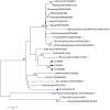
Figure 2. Phylogenetic analysis of the S genes from different PDCoV strains
The phylogenetic tree was constructed and analyzed using the neighbor-joining method of MEGA 6.0 software (http://www.megasoftware.net). Bootstrap values were calculated with 1000 replicates. Reference sequences obtained from GenBank are indicated by strain names and GenBank accession numbers. The S genes of PDCoV isolated from three swine farms in the present study are indicated with black triangles. The first strain of PDCoV S gene isolated by our lab is indicated with a blue triangle.
Figure 3
Figure 3. Intestinal changes in PDCoV infected piglets
(A) Piglets showed thin and transparent intestinal walls from colon to caecum (arrows). (B) Mesentery with spot hemorrhage (arrows). (C) Intestinal bleeding (arrows). (D) Stomach filled with curdled milk and accumulation of large amounts of yellow fluid in the jejunum lumen (arrows).
Figure 4
Figure 4. PDCoV distribution in various tissues
The virus copies (log10 GE/µg of total RNA) were mean virus copy of nine piglets. High PDCoV RNA copies were detected in ileum, inguinal lymph node, rectum and spleen. The highest PDCoV RNA copy was detected in ileum. Standard error bars are shown in each tissue.
Figure 5
Figure 5. Microscopic lesions and IHC staining
(A) H.E-stained jejunum of PDCoV-infected piglet with intestinal villus atrophy and acute diffuse jejunitis (original magnification ×40) (arrows). (B) H.E-stained jejunum tissue section of a control pig. (C) H.E-stained ileum of PDCoV infected piglet with intestinal acute, jejunitis diffuse cell proliferation and ileitis. (original magnification ×100). (arrows). (D) H.E-stained ileum tissue section of a control pig. (E) Section of jejunum of PDCoV-infected piglet, showing basal layer of intestine are positive for PDCoV RNA (original magnification ×400). (F) Section of ileum of PDCoV-infected piglet, with basal layer of intestine are positive for PDCoV RNA (original magnification ×400).

References

    1. Tohru S., Tomoyuki S., Naoto I.. et al. (2018) Genetic characterization and pathogenicity of Japanese porcine deltacoronavirus. Infect. Genet. Evol. 61, 176–182 10.1016/j.meegid.2018.03.030 - DOI - PMC - PubMed
    1. Woo P.C., Lau S.K., Lam C.S.. et al. (2012) Discovery of seven novel Mammalian and avian coronaviruses in the genus deltacoronavirus supports bat coronaviruses as the gene source of alphacoronavirus and betacoronavirus and avian coronaviruses as the gene source of gammacoronavirus and deltacoronavirus. J. Virol. 86, 3995–4008 10.1128/JVI.06540-11 - DOI - PMC - PubMed
    1. Marthaler D., Raymond L., Jiang Y.. et al. (2014) Rapid detection, complete genome sequencing, and phylogenetic analysis of porcine deltacoronavirus. Emerg. Infect. Dis. 20, 1347–1350 10.3201/eid2008.140526 - DOI - PMC - PubMed
    1. Wang L., Byrum B. and Zhang Y. (2014) Detection and genetic characterization of deltacoronavirus in pigs, Ohio, USA, 2014. Emerg. Infect. Dis. 20, 1227–1230 10.3201/eid2007.140296 - DOI - PMC - PubMed
    1. Lee S. and Lee C. (2014) Complete genome characterization of Korean porcine deltacoronavirus strain KOR/KNU14-04/2014. Genome Announc. 2, e01191–14 10.1128/genomeA.01191-14 - DOI - PMC - PubMed

Publication types

MeSH terms