. 2019 Sep 24;116(39):19530-19540.
doi: 10.1073/pnas.1908099116. Epub 2019 Sep 5.

Lgr5+ pericentral hepatocytes are self-maintained in normal liver regeneration and susceptible to hepatocarcinogenesis

Affiliations

Lgr5+ pericentral hepatocytes are self-maintained in normal liver regeneration and susceptible to hepatocarcinogenesis

Chow Hiang Ang et al. Proc Natl Acad Sci U S A. .

Abstract

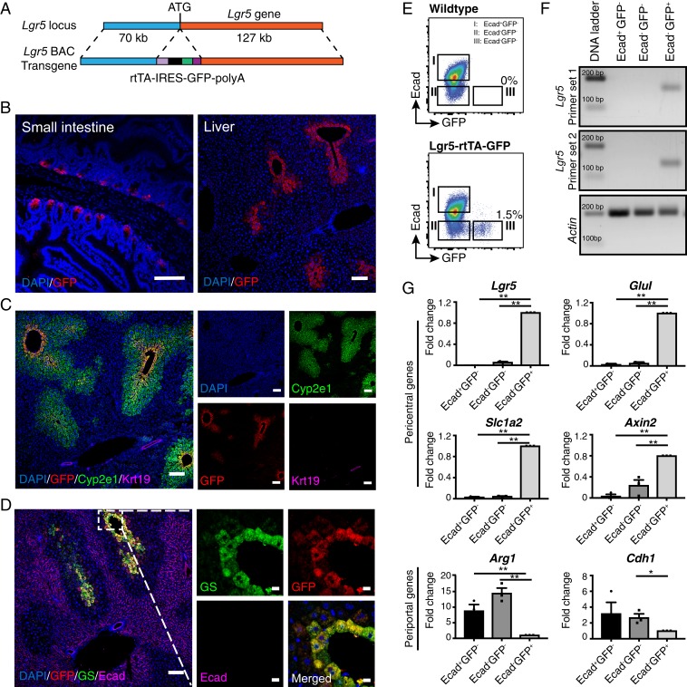
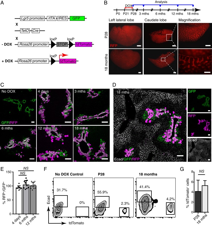
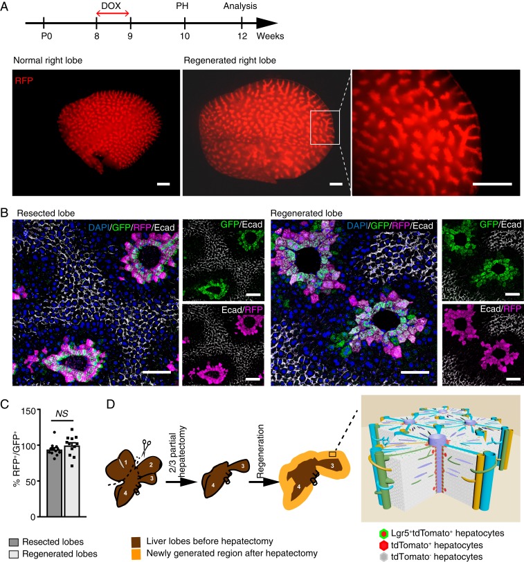
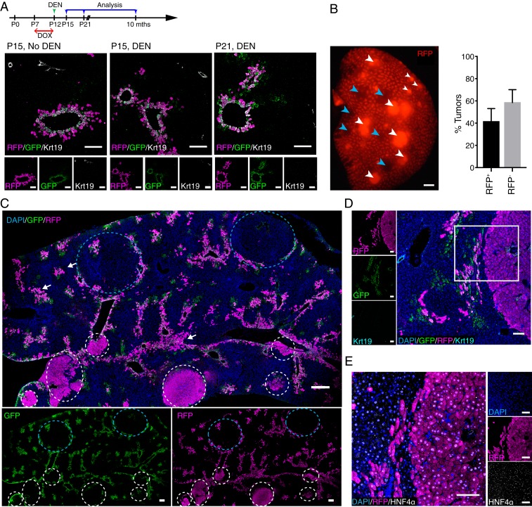
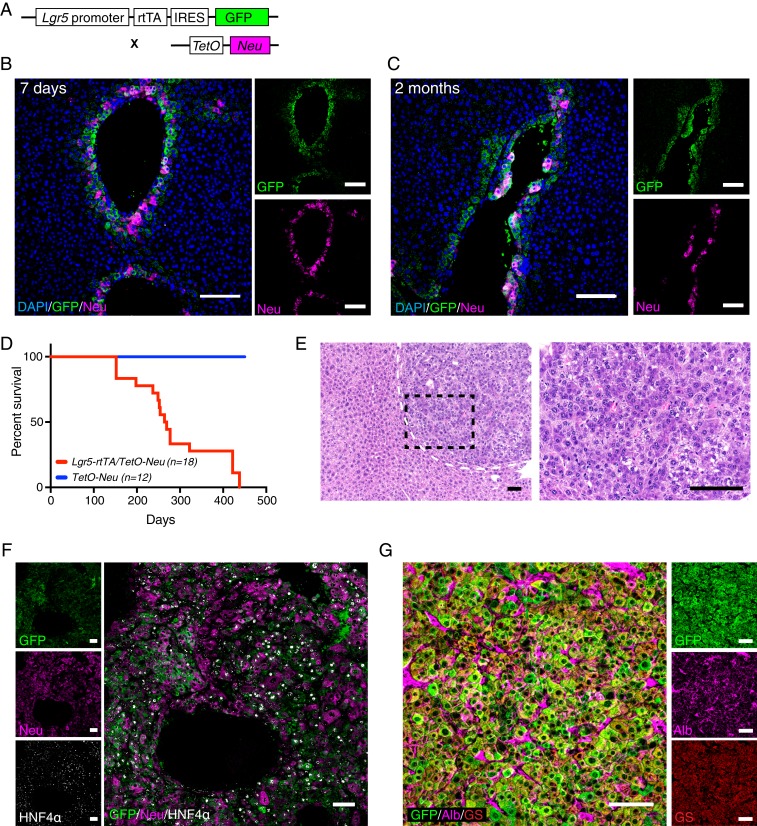
Emerging evidence suggests that hepatocytes are primarily maintained by self-renewal during normal liver homeostasis, as well as in response to a wide variety of hepatic injuries. However, how hepatocytes in distinct anatomic locations within the liver lobule are replenished under homeostasis and injury-induced regeneration remains elusive. Using a newly developed bacterial artificial chromosome (BAC)-transgenic mouse model, we demonstrate that Lgr5 expression in the liver is restricted to a unique subset of hepatocytes most adjacent to the central veins. Genetic lineage tracing revealed that pericentral Lgr5+ hepatocytes have a long lifespan and mainly contribute to their own lineage maintenance during postnatal liver development and homeostasis. Remarkably, these hepatocytes also fuel the regeneration of their own lineage during the massive and rapid regeneration process following two-thirds partial hepatectomy. Moreover, Lgr5+ hepatocytes are found to be the main cellular origin of diethylnitrosamine (DEN)-induced hepatocellular carcinoma (HCC) and are highly susceptible to neoplastic transformation triggered by activation of Erbb pathway. Our findings establish an unexpected self-maintaining mode for a defined subset of hepatocytes during liver homeostasis and regeneration, and identify Lgr5+ pericentral hepatocytes as major cells of origin in HCC development.

Keywords: Lgr5; hepatocellular carcinoma; hepatocyte; lineage tracing; liver regeneration.


Conflict of interest statement

The authors declare no conflict of interest.

Figures

Fig. 1.
Fig. 1.
Lgr5 is expressed in a small subset of hepatocytes mostly adjacent to the central veins. (A) Schematic illustration of the transgene structure for generation of Lgr5-rtTA-IRES-GFP mouse model. The rtTA-IRES-GFP-polyA cassette was inserted in frame to replace the ATG start codon of Lgr5 gene in a BAC DNA clone containing the Lgr5 gene locus. (B) Representative confocal images showing expression of GFP in small intestine (Left) and liver (Right) of adult Lgr5-rtTA-IRES-GFP transgenic mice. (C) Representative confocal images showing costaining of GFP with the pericentral hepatocyte marker Cyp2e1 and bile duct marker Krt19. (D) Representative confocal images depicting that the Lgr5-GFP+ population largely overlaps with the GS-expressing hepatocytes at the thin rim surrounding the central veins and is mutually exclusive to the cells marked by Ecad. (E) Representative FACS plots showing mutual exclusive presence of Lgr5-GFP+ hepatocytes and Ecad+ hepatocytes in adult WT and Lgr5-rtTA-IRES-GFP transgenic mice. (F) Representative gel images showing the expression of endogenous Lgr5 was restricted in the Lgr5-GFP+ hepatocytes. Three subsets of hepatocytes defined by Lgr5-GFP and Ecad expression were FACS sorted and two independent sets of primers targeting different exons of the Lgr5 gene were used for PCR amplification. (G) qPCR analysis of expression of periportal and pericentral genes in the three subsets of hepatocytes defined by Lgr5-GFP and Ecad expression. FACS isolated hepatocytes for each subpopulation were pooled from three mice to procure sufficient total RNA for qPCR analysis. Three independent experiments were conducted for qPCR analyses. *P < 0.05; **P < 0.01. Error bars represent mean ± SEM. (Scale bars: B, C, and D, Left, 100 μm; D, Right, 10 μm.)
Fig. 2.
Fig. 2.
Lgr5+ hepatocytes are long-lived and maintain their own lineage under homeostasis condition. (A) Schematic illustration of the genetic Lgr5-rtTA/TetO-Cre/R26-tdTomato mouse model for lineage tracing. Lgr5-rtTA-IRES-GFP mice were crossed with TetO-Cre mice and R26-tdTomato reporter strain to generate doxycycline (DOX)-inducible triple transgenic model. (B) Schematic illustration showing the experimental strategy for doxycycline induction and time points of analysis in lineage tracing of Lgr5-rtTA/TetO-Cre/R26-tdTomato mice. Representative whole-mount fluorescence images of the left lateral and caudate liver lobes showing identical expression pattern of tdTomato in the mice harvested immediately after doxycycline induction and at 18 mo after treatment. (C) Confocal imaging analysis of Lgr5-rtTA/TetO-Cre/R26-tdTomato mice chased for 4 d and 3, 6, 12, and 18 mo. Liver sections were stained with GFP and RFP antibodies. Triple transgenic mice without doxycycline treatment were used as control (No DOX). At least three mice were used for each experimental condition. (D) Representative confocal images for costaining of GFP, RFP, and Ecad on the liver sections of Lgr5-rtTA/TetO-Cre/R26-tdTomato mice chased for 18 mo. The tdTomato+ hepatocytes are mutually exclusive with periportal Ecad+ hepatocytes, indicating zone-restriction of progeny of Lgr5+ cells. (E) Quantification of the percentage of RFP+ in proportion to GFP+ hepatocytes in mice traced for 4 d, 6 mo, and 12 mo after doxycycline induction. An average of 300 cells per site were counted. n = 10 sites on the liver from three mice. Error bars represent mean ± SEM, *P < 0.05 is calculated by using multiple t test. No significant expansion was observed in comparisons of 4 d versus 6 mo (P = 0.07) and 4 d versus 12 mo (P = 0.12). (F) Representative FACS analysis of total hepatocytes from the Lgr5-rtTA/TetO-Cre/R26-tdTomato mice harvested immediately after doxycycline induction and chased for 18 mo. Lgr5-rtTA/TetO-Cre/R26-tdTomato mice without doxycycline treatment (No DOX) showed no tdTomato+ hepatocytes. (G) Bar graph showing no significant changes in percentage of tdTomato+ cells detected by FACS analysis of total hepatocytes from the Lgr5-rtTA/TetO-Cre/R26-tdTomato mice harvested immediately after doxycycline induction at P28 and chased for 18 mo. Error bars represent mean ± SEM, *P < 0.05 is calculated by using multiple t test. (Scale bars: B, 200 μm; C and D, 100 μm.)
Fig. 3.
Fig. 3.
Lgr5-derived hepatocytes replenish their own lineage upon partial hepatectomy. (A) Experimental strategy for partial hepatectomy experiments performed on Lgr5-rtTA/TetO-Cre/R26-tdTomato mice. Representative whole-mount images of the right liver lobe from adult littermate control and Lgr5-rtTA/TetO-Cre/R26-tdTomato mice harvested at 2 wk after partial hepatectomy. n = 5 mice. (B) Representative confocal images showing similar immunofluorescence staining pattern of Lgr5-GFP+tdTomato+ population in the resected liver lobe in comparison to the regenerated lobe harvested from the same mouse. Lgr5-GFP+tdTomato+ population remained in the thin-rim surrounding the central veins after two-thirds partial hepatectomy. n = 5. (C) Quantification of the percentage of RFP+ in proportion to GFP+ hepatocytes on resected lobes and regenerated lobes. An average of 200 cells per site were counted. n = 12 sites on the liver from three mice. Error bars represent mean ± SEM, *P < 0.05 is calculated by using multiple t test. (D) Graphic illustration of two-thirds partial hepatectomy procedure and liver regeneration after resection. Left lateral (1) and median (2) lobes are resected, and caudate (3) and right (4) lobes expand to restore the liver mass after surgery. A model illustrates the regeneration of Lgr5+ hepatocytes upon 2/3 partial hepatectomy. Lgr5+ pericentral hepatocytes mainly maintain their own lineage (Lgr5-GFP+tdTomato+) along the central vein upon two-thirds partial hepatectomy and occationally give rise to heptocytes in close pericentral layers (Lgr5-GFPtdTomato+) within the liver lobule. (Scale bars: A, 200 μm; B, 100 μm.)
Fig. 4.
Fig. 4.
Lgr5+ hepatocytes are the primary cellular origin in DEN-induced HCC. (A) Experimental strategy for doxycycline (DOX) and DEN induction for Lgr5-rtTA/TetO-Cre/R26-tdTomato mice. Representative confocal images showing distribution of Lgr5-GFP+tdTomato+ around central veins at P15 without DEN (25 mg/kg) injection, and at P15 and P21, which were 3 d and 9 d after DEN injection, respectively. Dams were treated with doxycycline at P7–P12 of their litters to induce the labeling of tdTomato in Lgr5-expressing hepatocytes of young male pups. (B) Representative whole-mount fluorescence view of a tumorous liver lobe of Lgr5-rtTA/TetO-Cre/R26-tdTomato mouse harvested at 8 mo after DEN induction. tdTomato+ and tdTomato tumors are depicted by white and blue arrowheads, respectively. Approximately 40% of the tumors detected in the liver of triple transgenic mice showed native tdTomato+ (RFP+) fluorescence. n = 4 mice. (C) Representative confocal tile scan images of a tumorous liver lobe harvested from DEN-treated Lgr5-rtTA/TetO-Cre/R26-tdTomato mouse showing tdTomato+ (white circles) and tdTomato (blue circles) tumors. White arrows indicate possible tdTomato+ tumor initiation sites. All DEN-induced tumors were negative for GFP. n = 4 mice. (D and E) Representative confocal images showing that tdTomato+ tumors are GFP and Krt19-negative (D), and HNF4α-positive (E). (Scale bars: B and C, 200 μm; D and E, 100 μm.)
Fig. 5.
Fig. 5.
Expression of constitutively active Neu/Her2 mutant in Lgr5+ hepatocytes induces hepatocellular carcinoma. (A) Schematic illustration of doxycycline-inducible Lgr5-rtTA/TetO-Neu mouse model. Constant treatment of doxycycline allows continuous expression of Neu/Her2 in Lgr5+ hepatocytes. (B and C) Representative confocal images showing expression of GFP and Neu/Her2 on liver sections of mice treated with doxycycline diet for 7 d (B) and 2 mo (C). Neu/Her2 expression was turned on specifically in Lgr5-GFP+ hepatocytes as early as 7 d. (D) Tumor latency in doxycycline-induced Lgr5-rtTA/TetO-Neu transgenic mice. Tumors were detected as early as 4 mo after starting doxycycline treatment. No tumor was found in control TetO-Neu mice. (E) Representative images of H&E staining of liver tumors arisen in Lgr5-rtTA/TetO-Neu mice at low magnification (Left) and high magnification (Right). n = 18 mice. (F) Costaining of GFP, Neu/Her2, an HNF4α revealed that Lgr5-rtTA/TetO-Neu tumors are HNF4α+, and majority of the tumor cells retained expression of Lgr5-GFP and Neu/Her2 (n = 4). (G) Representative confocal image showing Lgr5-rtTA/TetO-Neu tumor cells express hepatocyte markers albumin and GS (n = 4 mice). (Scale bars: 100 μm.)

References

    1. Kopp J. L., Grompe M., Sander M., Stem cells versus plasticity in liver and pancreas regeneration. Nat. Cell Biol. 18, 238–245 (2016). - PubMed
    1. Miyajima A., Tanaka M., Itoh T., Stem/progenitor cells in liver development, homeostasis, regeneration, and reprogramming. Cell Stem Cell 14, 561–574 (2014). - PubMed
    1. Yanger K., et al. , Adult hepatocytes are generated by self-duplication rather than stem cell differentiation. Cell Stem Cell 15, 340–349 (2014). - PMC - PubMed
    1. Schaub J. R., Malato Y., Gormond C., Willenbring H., Evidence against a stem cell origin of new hepatocytes in a common mouse model of chronic liver injury. Cell Rep. 8, 933–939 (2014). - PMC - PubMed
    1. Tarlow B. D., et al. , Bipotential adult liver progenitors are derived from chronically injured mature hepatocytes. Cell Stem Cell 15, 605–618 (2014). - PMC - PubMed

Publication types

MeSH terms

LinkOut - more resources