. 2018 Apr 23;4(3):20180007.
doi: 10.1259/bjrcr.20180007. eCollection 2018 Mar.

Ectopic meningioma in a patient with neurofibromatosis Type 2: a case report and review of the literature

Affiliations

Ectopic meningioma in a patient with neurofibromatosis Type 2: a case report and review of the literature

Chun Qiu Su et al. BJR Case Rep. .

Abstract

Ectopic meningioma occurring in the region of parapharyngeal space is rare in clinical practice and brings great challenge in its diagnosis. This report details such a case in a 14-year-old girl with neurofibromatosis Type 2, which is a highly infrequent association. The clinical manifestations, imaging findings, and pathological manifestations are described, and the relevant literature is reviewed to highlight characteristic imaging findings of ectopic meningiomas.


Figures
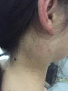
Figure 1.
Figure 1.
The patient, a 14-year-old girl, presented with a painless mass in the right retroauricular area.
Figure 2.
Figure 2.
Nasopharyngeal MRI examination reveals a large, well-circumscribed mass in the right parapharyngeal space. The mass is isohypointense on T 1WI (a) and slightly hyperintense on T 2WI (b); it is accompanied by multiple focal hypointensities that may represent calcification. Marked but heterogeneous enhancement was observed on axial post-contrast T 1WI after the administration of gadolinium (c). The sagittal post-contrast T 1WI (f) shows that the mass had invaded the carotid space—as well as the parotid and perivertebral space—and encased major carotid vessels. DWI (d) revealed a restriction of diffusion, with an average ADC value of 699.7 × 10⁻⁶ mm2 s 1 (e). ADC, apparent diffusion coefficient; DWI, diffusion-weighted imaging; T 1WI,T weighted imaging; T 2WI,T 2 weighted imaging.
Figure 3.
Figure 3.
An axial post-contrast T 1WI (a–c), reveals multiple nodular masses in the frontal region and the midline (black arrows). There were bilateral masses in the bilateral cerebellopontine angle, which broadened bilateral internal auditory canal. They showed significant contrast enhancement after gadolinium administration (white arrow). There were bilateral thickening C6-C7 spinal nerve root. These roots were isointense on both coronal T 1WI (f) and T 2WI (g). In addition, obvious nodular enhancement is seen on the coronal post-contrast T 1WI (h) (red arrows). The coronal post-contrast T 1WI shows involvement of the right hypoglossal canal (yellow arrows). T 1WI,T weighted imaging; T 2WI,T 2 weighted imaging.
Figure 4.
Figure 4.
(a) H-E staining shows psamomma bodies in sheets of cells with a meningothelial pattern (H-E;×200). (b) Immunohistochemistry shows positive staining for EMA. (Magnification × 100). EMA, epithelial membrane antigen; H-E, haematoxylin and eosin.

References

    1. Rushing EJ, Bouffard JP, McCall S, Olsen C, Mena H, Sandberg GD, et al. . Primary extracranial meningiomas: an analysis of 146 cases. Head Neck Pathol 2009; 3: 116–30. doi: 10.1007/s12105-009-0118-1 - DOI - PMC - PubMed
    1. Verma SK, Satyarthee G, Borkar SA, Singh M, Sharma BS. Orbital roof intradiploic meningioma in a 16-year-old girl. J Pediatr Neurosci 2015; 10: 51–4. doi: 10.4103/1817-1745.154342 - DOI - PMC - PubMed
    1. Sanei MH, Rabiei S, Eftekhari M, Jafari HR. Ectopic meningioma (hamartoma) of the middle ear: a challenging case in frozen section. Otol Neurotol 2014; 35: e231–2. doi: 10.1097/MAO.0000000000000404 - DOI - PubMed
    1. Ma C, Li X, Li Y, Qu X. Primary Ectopic Meningioma of the Tongue: Case Report and Review of the Literature. J Oral Maxillofac Surg 2016; 74: 2216–28. doi: 10.1016/j.joms.2016.04.023 - DOI - PubMed
    1. Lu C, Hu X, Xu M, Mao W, Yang H, Wang Z, et al. . Posterior mediastinal ectopic meningioma: a case report. World J Surg Oncol 2015; 13: 156. doi: 10.1186/s12957-015-0581-y - DOI - PMC - PubMed