Neurodegeneration with Brain Iron Accumulation: Two Additional Cases with Dystonic Opisthotonus
- PMID: 31489256
- PMCID: PMC6707210
- DOI: 10.7916/tohm.v0.683
Neurodegeneration with Brain Iron Accumulation: Two Additional Cases with Dystonic Opisthotonus
Abstract
Background: Specific phenomenology and pattern of involvement in movement disorders point toward a probable clinical diagnosis. For example, forehead chorea usually suggests Huntington's disease; feeding dystonia suggests neuroacanthocytosis and risus sardonicus is commonly seen in Wilson's disease. Dystonic opisthotonus has been described as a characteristic feature of neurodegeneration with brain iron accumulation (NBIA) related to PANK2 and PLA2G6 mutations.
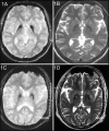
Case report: We describe two additional patients in their 30s with severe extensor truncal dystonia causing opisthotonic posturing in whom evaluation revealed the diagnosis of NBIA confirmed by genetic testing.
Discussion: Dystonic opisthotonus may be more common in NBIA than it is reported and its presence especially in a young patient should alert the neurologists to a possibility of probable NBIA.
Keywords: Opisthotonus; botulinum toxin; dystonia; genetics; neurodegeneration with brain iron accumulation; phenomenology; secondary.
Conflict of interest statement
Funding: None. Conflicts of Interest: The authors report no conflicts of interest. Ethics Statement: All patients that appear on video have provided written informed consent; authorization for the videotaping and for publication of the videotape was provided.
Figures
References
Publication types
MeSH terms
Supplementary concepts
LinkOut - more resources
Full Text Sources
Medical


