. 2019 Aug 28;5(8):eaaw9298.
doi: 10.1126/sciadv.aaw9298. eCollection 2019 Aug.

Cushing's syndrome driver mutation disrupts protein kinase A allosteric network, altering both regulation and substrate specificity

Affiliations

Cushing's syndrome driver mutation disrupts protein kinase A allosteric network, altering both regulation and substrate specificity

Caitlin Walker et al. Sci Adv. .

Abstract

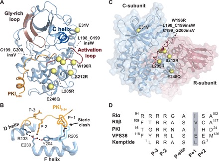
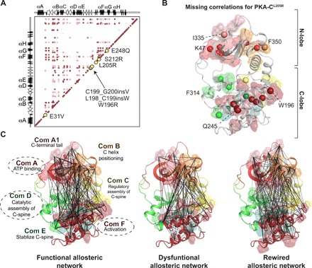
Genetic alterations in the PRKACA gene coding for the catalytic α subunit of the cAMP-dependent protein kinase A (PKA-C) are linked to cortisol-secreting adrenocortical adenomas, resulting in Cushing's syndrome. Among those, a single mutation (L205R) has been found in up to 67% of patients. Because the x-ray structures of the wild-type and mutant kinases are essentially identical, the mechanism explaining aberrant function of this mutant remains under active debate. Using NMR spectroscopy, thermodynamics, kinetic assays, and molecular dynamics simulations, we found that this single mutation causes global changes in the enzyme, disrupting the intramolecular allosteric network and eliciting losses in nucleotide/pseudo-substrate binding cooperativity. Remarkably, by rewiring its internal allosteric network, PKA-CL205R is able to bind and phosphorylate non-canonical substrates, explaining its changes in substrate specificity. Both the lack of regulation and change in substrate specificity reveal the complex role of this mutated kinase in the formation of cortisol-secreting adrenocortical adenomas.


Figures

Fig. 1
Fig. 1. Architecture of PKA-C and locations of the Cushing’s syndrome mutations.
(A) Structure of PKA-C [Protein Data Bank (PDB) ID: 1ATP] in complex with pseudo-substrate PKI depicting the location of Cushing’s mutations (yellow spheres) in relation to structural elements of the kinase. The E248Q mutation includes an additional deletion (del243-247), and the S212R mutation includes an insertion (insIILR) not depicted. (B) X-ray structure of PKA-CL205R (PDB ID: 4WB6) with the overlay of PKI5–24 (PKIWT from PDB ID: 1ATP) describing the architecture of the peptide binding site and steric clash between kinase (PKA-CL205R) and pseudo-substrate. (C) Structure of the R/C complex (PDB ID: 2QCS) depicting locations of Cushing’s mutations in relation to the pseudo-substrate inhibitory sequence of the R-subunit. (D) Primary sequence comparison of common regulators (RIα, RIIβ, and PKI) and peptide substrates of the catalytic subunit (Kemptide and VPS36).
Fig. 2
Fig. 2. Allosteric network of interactions observed upon pseudo-substrate binding.
The CHESCA correlation matrix for (A) PKA-CWT upon binding PKI and (B) PKA-CL205R upon binding PKI. (C) Correlations corresponding to the binding of PKI to PKA-CWT plotted on the structure of PKA-C. Residues that are commonly correlated for both PKA-CWT and PKA-CL205R are highlighted. (D) Correlations corresponding to the binding of PKI to PKA-CL205R plotted on the structure of PKA-C. Specific residues that are correlated for only PKA-CL205R are highlighted. Only correlations with rij > 0.98 are shown throughout.
Fig. 3
Fig. 3. Rewiring of the allosteric network of PKA-CL205R upon binding VPS36.
(A) Steady-state phosphorylation kinetics of Kemptide and VPS36 peptides for PKA-CWT and PKA-CL205R. Corresponding values can be found in table S2. (B) CONCISE analysis on the apo, ATPγN, ATPγN/PKI, and ATPγN/VPS36 states of PKA-CWT and PKA-CL205R. (C) The CHESCA correlation matrix for PKA-CWT upon binding VPS36. (D) The CHESCA correlation matrix for PKA-CL205R upon binding VPS36.
Fig. 4
Fig. 4. Conformational dynamics of the activation loop of PKA-CWT and PKA-CL205R upon binding substrate.
(A) Distinct opening-closing motions of the Gly-rich loop highlighting the hindered conformation that occludes the entering of ATP in PKA-CL205R. (B) Probability density describing the conformation of the activation loop in response to different substrates and pseudo-substrates. (C) [1H, 15N]-TROSY-HSQC spectra showing the backbone amide chemical shift changes of the W196 indole amide (located on the activation loop) in response to binding PKI or VPS36. (D) X-ray structure of PKA-CWT (gray) (PDB ID: 1ATP) with the overlay PKA-CL205R in complex with PKI (light blue) (PDB ID: 4WB6) describing the architecture of the peptide binding site and activation loop flip. (E) X-ray structure of PKA-CWT (gray) (PDB ID: 1ATP) with the overlay PKA-CL205R in complex with VPS36 (light blue) (MD simulations) describing the architecture of the peptide binding site and activation loop flip.
Fig. 5
Fig. 5. Cushing’s mutations are located in allosteric nodes identified via CHESCA.
(A) Correlation matrix of PKA-CWT when bound to PKI emphasizing locations of Cushing’s mutation in relation to allosteric nodes. (B) Missing correlations for PKA-CL205R upon binding PKI colored according to the community map of PKA-CWT. (C) CHESCA correlation matrices for PKA-CWT + PKI, PKA-CL205R + PKI, and PKA-CL205R + VPS36, respectively, plotted on the structure of PKA-C with colors specific to the community map analyses completed previously for PKA-CWT (35). Specific communities are emphasized to highlight elements that experience the most dramatic changes in the number and extent of chemical shift covariance for PKA-CL205R upon binding PKI and VPS36.
Fig. 6
Fig. 6. Schematic of the energy landscape for PKA-CWT and PKA-CL205R, combining thermodynamics and MD simulations data.
Relative free energy for the binding of ATPγN, PKI, and VPS36 to PKA-CWT and PKA-CL205R derived from ITC data. (1) Apo PKA-CL205R samples mostly uncommitted states, with the Gly-rich loop partially occluded and the αC helix turned outward. (2) Binary PKA-CL205R features a wired allosteric network for substrate binding, i.e., committed state. (3) Ternary PKA-CL205R complex with lower affinity for PKI (i.e., higher free energy relative to PKA-CWT). The conformation of the activation loop of PKA-CL205R is in equilibrium between an unflipped and a sparsely populated flipped conformation. (4) PKA-CL205R/ATPγN/VPS36 ternary complex features a flipped conformation of the activation loop with the electrostatic interactions between E86 and R194.

References

    1. Hunter T., Why nature chose phosphate to modify proteins. Philos. Trans. R. Soc. Lond. B Biol. Sci. 367, 2513–2516 (2012). - PMC - PubMed
    1. Lodish M., Stratakis C. A., A genetic and molecular update on adrenocortical causes of Cushing syndrome. Nat. Rev. Endocrinol. 12, 255–262 (2016). - PubMed
    1. Cao Y., He M., Gao Z., Peng Y., Li Y., Li L., Zhou W., Li X., Zhong X., Lei Y., Su T., Wang H., Jiang Y., Yang L., Wei W., Yang X., Jiang X., Liu L., He J., Ye J., Wei Q., Li Y., Wang W., Wang J., Ning G., Activating hotspot L205r mutation in PRKACA and adrenal Cushing's syndrome. Science 344, 913–917 (2014). - PubMed
    1. Sato Y., Maekawa S., Ishii R., Sanada M., Morikawa T., Shiraishi Y., Yoshida K., Nagata Y., Sato-Otsubo A., Yoshizato T., Suzuki H., Shiozawa Y., Kataoka K., Kon A., Aoki K., Chiba K., Tanaka H., Kume H., Miyano S., Fukayama M., Nureki O., Homma Y., Ogawa S., Recurrent somatic mutations underlie corticotropin-independent Cushing’s syndrome. Science 344, 917–920 (2014). - PubMed
    1. Goh G., Scholl U. I., Healy J. M., Choi M., Prasad M. L., Nelson-Williams C., Kunstman J. W., Korah R., Suttorp A.-C., Dietrich D., Haase M., Willenberg H. S., Stålberg P., Hellman P., Åkerström G., Björklund P., Carling T., Lifton R. P., Recurrent activating mutation in PRKACA in cortisol-producing adrenal tumors. Nat. Genet. 46, 613–617 (2014). - PMC - PubMed

Publication types

MeSH terms

Substances