. 2019 Aug 28;5(8):eaax2476.
doi: 10.1126/sciadv.aax2476. eCollection 2019 Aug.

Combinatorial morphogenetic and mechanical cues to mimic bone development for defect repair

Affiliations

Combinatorial morphogenetic and mechanical cues to mimic bone development for defect repair

S Herberg et al. Sci Adv. .

Abstract

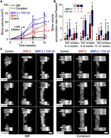
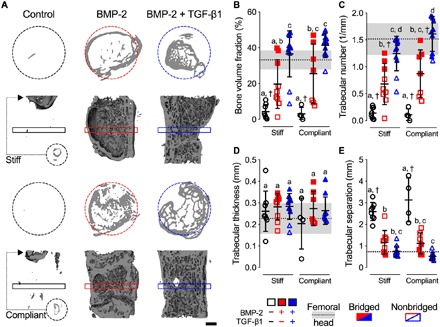
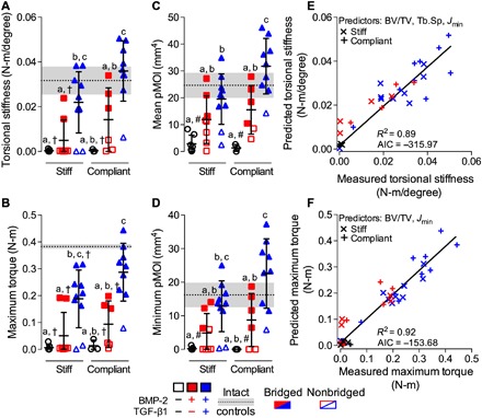
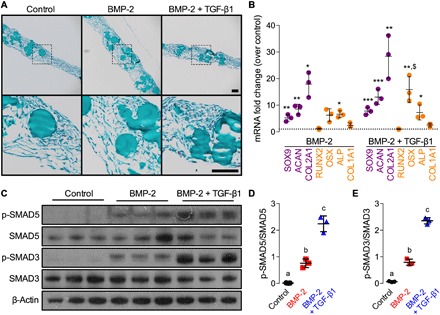
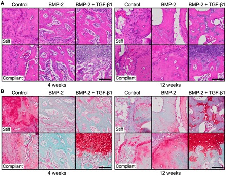
Endochondral ossification during long bone development and natural fracture healing initiates by mesenchymal cell condensation, directed by local morphogen signals and mechanical cues. Here, we aimed to mimic development for regeneration of large bone defects. We hypothesized that engineered human mesenchymal condensations presenting transforming growth factor-β1 (TGF-β1) and/or bone morphogenetic protein-2 (BMP-2) from encapsulated microparticles promotes endochondral defect regeneration contingent on in vivo mechanical cues. Mesenchymal condensations induced bone formation dependent on morphogen presentation, with BMP-2 + TGF-β1 fully restoring mechanical function. Delayed in vivo ambulatory loading significantly enhanced the bone formation rate in the dual morphogen group. In vitro, BMP-2 or BMP-2 + TGF-β1 initiated robust endochondral lineage commitment. In vivo, however, extensive cartilage formation was evident predominantly in the BMP-2 + TGF-β1 group, enhanced by mechanical loading. Together, this study demonstrates a biomimetic template for recapitulating developmental morphogenic and mechanical cues in vivo for tissue engineering.


Figures

Fig. 1
Fig. 1. Effects of morphogen priming of engineered mesenchymal condensations and in vivo mechanical loading on longitudinal bone formation and bone accumulation rate.
(A) Longitudinal quantification of bone volume at weeks 4, 8, and 12 by in vivo microCT (n = 4 to 11 per group). Data are shown as means ± SD. (B) Bone volume accumulation rate, defined as bone volume accrual over each 4-week interval. Box plots display median as horizontal line, mean as +, interquartile range as boxes, and minimum/maximum range as whiskers. (C) Representative three-dimensional (3D) microCT reconstructions showing bone formation per group over time. Representative samples were selected on the basis of mean bone volume at week 12. Scale bars, 3 mm. Comparisons between groups were evaluated by two-way repeated measures analysis of variance (ANOVA) with Tukey’s post hoc tests. Repeated significance indicator letters (a, b, and c) signify P > 0.05, while groups with distinct indicators signify P < 0.05 at each time point. Comparisons between time points were not assessed.
Fig. 2
Fig. 2. Effects of morphogen priming of engineered mesenchymal condensations and in vivo mechanical loading on new bone quantity and architecture.
(A) Representative 3D microCT reconstructions, with bone formation illustrated at mid-shaft transverse (top) and sagittal (bottom) sections at week 12, selected on the basis of mean bone volume. Dashed circles show 5-mm defect region. Rectangular boxes illustrate transverse cutting planes. Note that, due to minimal bone regeneration, additional transverse sections for stiff and compliant no growth factor controls were derived from the proximal end of the defect (small dashed circles and arrows). Scale bar, 1 mm. (B) Morphometric analysis of bone volume fraction, (C) trabecular number, (D) trabecular thickness, and (E) trabecular separation at week 12 (n = 4 to 11 per group), shown with corresponding measured parameters of femoral head trabecular bone (n = 3; dotted lines with gray shading: means ± SD; †P < 0.05 versus femoral head). Individual data points are shown as means ± SD. Comparisons between groups were evaluated by two-way ANOVA with Tukey’s post hoc tests. Repeated significance indicator letters (a, b, and c) signify P > 0.05, while groups with distinct indicators signify P < 0.05.
Fig. 3
Fig. 3. Effects of morphogen priming of engineered mesenchymal condensations and in vivo mechanical loading on functional defect regeneration.
(A) Torsional stiffness, (B) maximum torque at failure, (C) mean pMOI, and (D) minimum pMOI. Best subset regression analysis (R2) with lowest Akaike’s information criterion (AIC) value for measured and predicted (E) torsional stiffness and (F) maximum torque at failure indicating significant contributions of bone volume fraction (BV/TV), trabecular separation (Tb.Sp), and minimum pMOI (Jmin). Individual data points are shown as means ± SD (n = 3 to 10 per group). Comparisons between groups were evaluated by two-way ANOVA with Tukey’s post hoc tests. Repeated significance indicator letters (a, b, and c) signify P > 0.05, while groups with distinct indicators signify P < 0.05. Biomechanical and structural parameters are shown with age-matched intact bone properties, with pMOI obtained from the same mid-shaft region of interest (ROI) as used for the defects (n = 3; dotted lines with gray shading: means ± SD; †P < 0.05 and #P < 0.05 versus intact bone).
Fig. 4
Fig. 4. Effects of morphogen priming of engineered mesenchymal condensations on in vitro chondrogenic lineage specification at the time of implantation.
Histological Safranin-O/Fast green staining of representative microparticle-containing hMSC sheets at the time of implantation (2 days; n = 3 per group). Scale bars, 100 μm (top, x10; bottom, x40 magnification of dotted squares). (B) Normalized mRNA fold change over control of key chondrogenic or osteogenic markers by quantitative reverse transcription polymerase chain reaction (qRT-PCR; n = 3 per group; *P < 0.05, **P < 0.01, ***P < 0.001 versus empty/control; $P < 0.05 versus BMP-2–containing hMSC sheets). (C) Representative immunoblots and (D) relative quantification of phosphorylated SMAD5 (p-SMAD5)/SMAD5 and (E) p-SMAD3/SMAD3 in lysates of day 2 hMSC sheets (n = 3 per group). β-Actin served as the loading control. Individual data points are shown as means ± SD. Comparisons between groups were evaluated by one-way ANOVA with Tukey’s post hoc tests. Repeated significance indicator letters (a, b, and c) signify P > 0.05, while groups with distinct indicators signify P < 0.05.
Fig. 5
Fig. 5. Effects of morphogen priming of engineered mesenchymal condensations and in vivo mechanical loading on tissue-level bone regeneration.
Representative histological (A) hematoxylin and eosin and (B) Safranin-O/Fast green staining of defect tissue at week 4 (left) and week 12 (right), with stiff (top) and compliant fixation (bottom), selected on the basis of mean bone volume. Scale bars, 100 μm (×40).

References

    1. Lefebvre V., Bhattaram P., Vertebrate skeletogenesis. Curr. Top. Dev. Biol. 90, 291–317 (2010). - PMC - PubMed
    1. Gerber H. P., Vu T. H., Ryan A. M., Kowalski J., Werb Z., Ferrara N., VEGF couples hypertrophic cartilage remodeling, ossification and angiogenesis during endochondral bone formation. Nat. Med. 5, 623–628 (1999). - PubMed
    1. Hosseini A., Hogg D. A., The effects of paralysis on skeletal development in the chick embryo. I. General effects. J. Anat. 177, 159–168 (1991). - PMC - PubMed
    1. Verbruggen S. W., Kainz B., Shelmerdine S. C., Hajnal J. V., Rutherford M. A., Arthurs O. J., Phillips A. T. M., Nowlan N. C., Stresses and strains on the human fetal skeleton during development. J. R. Soc. Interface 15, 20170593 (2018). - PMC - PubMed
    1. Carter D. R., Van Der Meulen M. C., Beaupré G. S., Mechanical factors in bone growth and development. Bone 18, 5S–10S (1996). - PubMed

Publication types

Substances

LinkOut - more resources