. 2019 Nov;23(11):7268-7278.
doi: 10.1111/jcmm.14555. Epub 2019 Sep 5.

Exosomes from miR-20b-3p-overexpressing stromal cells ameliorate calcium oxalate deposition in rat kidney

Affiliations

Exosomes from miR-20b-3p-overexpressing stromal cells ameliorate calcium oxalate deposition in rat kidney

Jing Shi et al. J Cell Mol Med. 2019 Nov.

Abstract

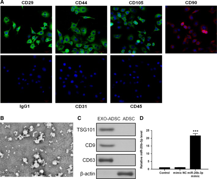
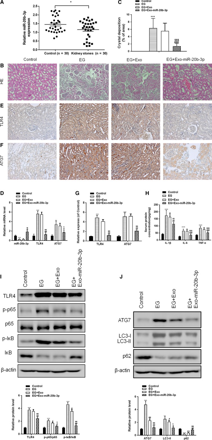
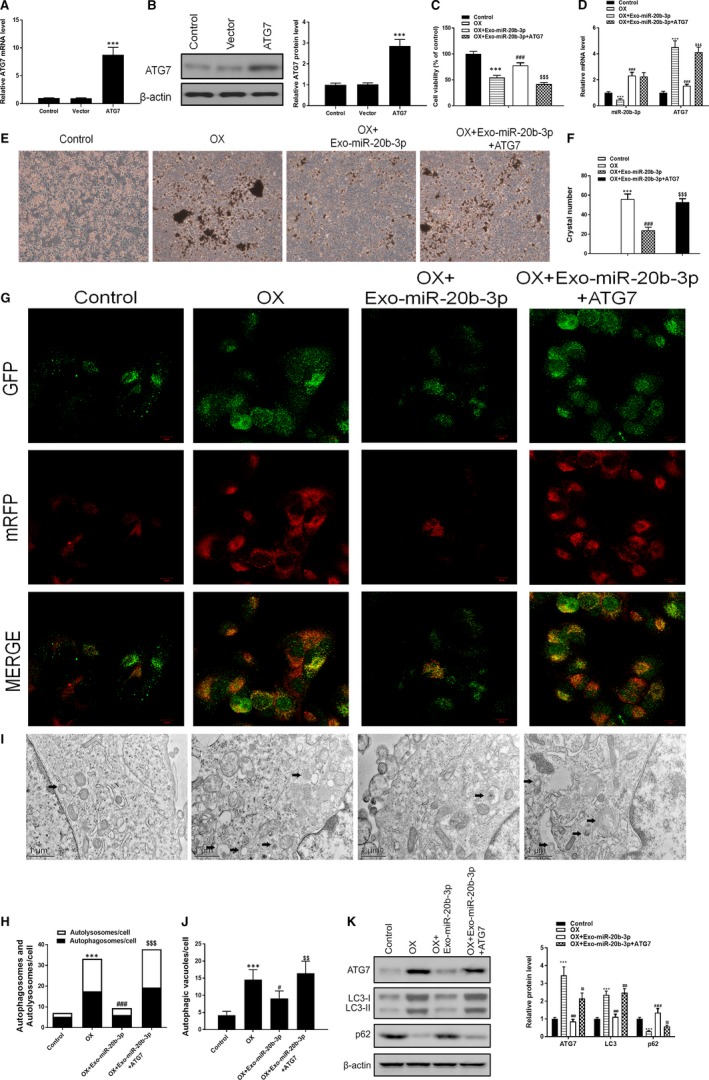
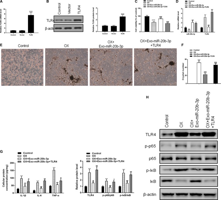
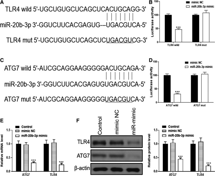
Hyperoxaluria-induced calcium oxalate (CaOx) deposition is the key factor in kidney stone formation, for which adipose-derived stromal cells (ADSCs) have been used as a therapeutic treatment. Studies revealed that miR-20b-3p is down-regulated in hypercalciuric stone-forming rat kidney. To investigate whether ADSC-derived miR-20b-3p-enriched exosomes protect against kidney stones, an ethylene glycol (EG)-induced hyperoxaluria rat model and an in vitro model of oxalate-induced NRK-52E cells were established to explore the protective mechanism of miR-20b-3p. The results showed that miR-20b-3p levels were decreased following hyperoxaluria in the urine of patients and in kidney tissues from animal models. Furthermore, treatment with miR-20b-3p-enriched exosomes from ADSCs protected EG-induced hyperoxaluria rats, and cell experiments confirmed that co-culture with miR-20b-3p-enriched exosomes alleviated oxalate-induced cell autophagy and the inflammatory response by inhibiting ATG7 and TLR4. In conclusion, ADSC-derived miR-20b-3p-enriched exosomes protected against kidney stones by suppressing autophagy and inflammatory responses.

Keywords: calcium oxalate deposition; exosomes; miR-20b-3p; microRNAs; stromal cells.


Conflict of interest statement

All authors declare no conflicts of interest.

Figures

Figure 1
Figure 1
Identification of adipose‐derived stromal cells (ADSCs) and exosomes. A, Assessment of cell surface markers by immunofluorescence staining. ADSCs were positive for mesenchymal stem cell markers CD29, CD44, CD90 and CD105, but negative for endothelial markers CD31 and CD45. Negative isotype controls are shown. Scale bars = 100 mm. B, Transmission electron microscopy image of exosomes. C, Western blots using CD63 and TSG101 as markers of ADSC‐derived exosomes. D, Reverse transcription PCR detection showing expression of miR‐20b‐3p in exosomes after transfection. Data are presented as the mean ± SD (n = 3). ***P < .001 vs control group
Figure 2
Figure 2
Treatment with adipose‐derived stromal cell‐derived miR‐20b‐3p‐enriched exosomes alleviates ethylene glycol (EG)‐induced kidney CaOx crystal deposition in rats. (A) RT‐PCR analyses of miR‐20b‐3p levels in the urine of kidney stone patients. (B and C) Photomicrographs of kidney sections from rats obtained under dark field illumination with polarized light. Retained crystals exhibit strong birefringence (200× magnification). (D) Levels of miR‐20b‐3p, ATG7 and TLR4 determined by RT‐PCR. Levels of ATG7 (E) and TLR4 (F) in kidney tissue were assessed by immunohistochemical staining and quantified (G). (H) Rat serum protein concentrations for IL‐1β, IL‐6 and TNF‐α determined by ELISA. (I) Inflammation‐related proteins TLR4, p65 and IkB determined by Western blotting and quantified. (J) Autophagy‐related proteins Atg7, P62 and LC3 determined by Western blotting and quantified. Data are presented as the mean ± SD (n = 6). **P < .01 and ***P < .001 vs control group; #P < .05, ##P < .01 and ###P < .001 vs EG group; $P < .05, $$P < .01 and $$$P < .001 vs EG + Exo group
Figure 3
Figure 3
Co‐culturing with miR‐20b‐3p‐enriched exosomes alleviates oxalate‐induced injury in NRK‐52E cells by inhibiting autophagy via ATG7. A and B, ATG7 levels in NRK‐52E cells after ATG7 overexpression for 48 h determined by RT‐PCR and Western blotting. C, Cell viability assessed by MTT assay. D, Levels of miR‐20b‐3p and ATG7 determined by RT‐PCR. (E and F) Crystal cell adhesion assay determination and quantification. G and H, Representative images of immunofluorescence staining of mRFP‐GFP‐LC3 in NRK‐52E cells. Representative profiles of autophagosomes (RFP + GFP+dots) and autolysosomes (RFP + GFP‐dots) per cell section assessed by confocal microscopy are shown and were quantified. I and J, Autophagic vacuoles (autophagosomes) were detected by transmission electron microscopy (TEM). Representative TEM images are shown and typical autophagosomes are marked with black arrows. The number of autophagosomes per cell was calculated by counting the number of double‐membrane organelles in 10 cells. K, Autophagy‐related proteins Atg7, P62 and LC3 determined by Western blotting and quantified. Data are presented as the mean ± SD (n = 3). **P < .01 and ***P < .001 vs control group; #P < .05, ##P < .01 and ###P < .001 vs OX group; $P < .05, $$P < .01 and $$$P < .001 vs OX + Exo‐miR‐20b‐3p group
Figure 4
Figure 4
Co‐culturing with miR‐20b‐3p‐enriched exosomes alleviates oxalate‐induced injury in NRK‐52E cells by inhibiting inflammatory responses via TLR4. A and B, TLR4 levels in NRK‐52E cells after TLR4 overexpression for 48 h determined by RT‐PCR and Western blotting. C, Cell viability assessed by MTT assay. D, Levels of miR‐20b‐3p and ATG7 determined by RT‐PCR. E and F, Crystal cell adhesion assay determination and quantitation. G, Cellular protein concentrations for IL‐1β, IL‐6 and TNF‐α determined by ELISA. H, Inflammation‐related proteins TLR4, p65 and IkB determined by Western blotting and quantified. Data are presented as the mean ± SD (n = 3). **P < .01 and ***P < .001 vs control group; #P < .05, ##P < .01 and ###P < .001 vs OX group; $P < .05, $$P < .01 and $$$P < .001 vs OX + Exo‐miR‐20b‐3p group
Figure 5
Figure 5
Both ATG7 and TLR4 are targets of miR‐20b‐3p. A and B, Complementary sequences between miR‐20b‐3p and the 3′‐untranslated region (UTR) of wild‐type or mutated version of ATG7 and TLR4 were obtained using publicly available algorithms. C and D, The 3′‐UTR of ATG7 and TLR4 was fused to the luciferase coding region and co‐transfected into NRK‐52E cells with miR‐20b‐3p mimic to confirm that miR‐20b‐3p is a target of miR‐20b‐3p. Relative luciferase activity was determined at 48 h after transfection. E and F, Levels of TLR4 and ATG7 determined by Western blotting and RT‐PCR. Data are presented as the mean ± SD (n = 3). ***P < .001 vs mimic NC

Comment in

Similar articles

Cited by

References

    1. Kusmartsev S, Dominguez‐Gutierrez PR, Canales BK, Bird VG, Vieweg J, Khan SR. Calcium oxalate stone fragment and crystal phagocytosis by human macrophages. J Urol. 2016;195:1143‐1151. - PMC - PubMed
    1. Zeng G, Mai Z, Xia S, et al. Prevalence of kidney stones in China: an ultrasonography based cross‐sectional study. BJU Int. 2017;120:109‐116. - PubMed
    1. Evan AP, Worcester EM, Coe FL, Williams J, Lingeman JE. Mechanisms of human kidney stone formation. Urolithiasis. 2015;43(Suppl 1):19‐32. - PMC - PubMed
    1. Worcester EM, Coe FL. Clinical practice. Calcium kidney stones. N Engl J Med. 2010;363:954‐963. - PMC - PubMed
    1. Burgos‐Silva M, Semedo‐Kuriki P, Donizetti‐Oliveira C, et al. Adipose tissue‐derived stem cells reduce acute and chronic kidney damage in mice. PLoS ONE. 2015;10:e0142183. - PMC - PubMed

Publication types

MeSH terms