. 2019;20(10):816-827.
doi: 10.1631/jzus.B1900071.

Catalpol ameliorates LPS-induced endometritis by inhibiting inflammation and TLR4/NF-κB signaling

Affiliations

Catalpol ameliorates LPS-induced endometritis by inhibiting inflammation and TLR4/NF-κB signaling

Hua Zhang et al. J Zhejiang Univ Sci B. 2019.

Erratum in

Abstract

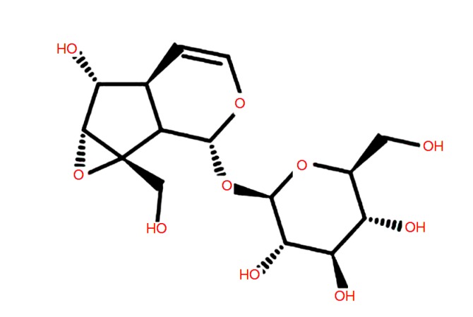
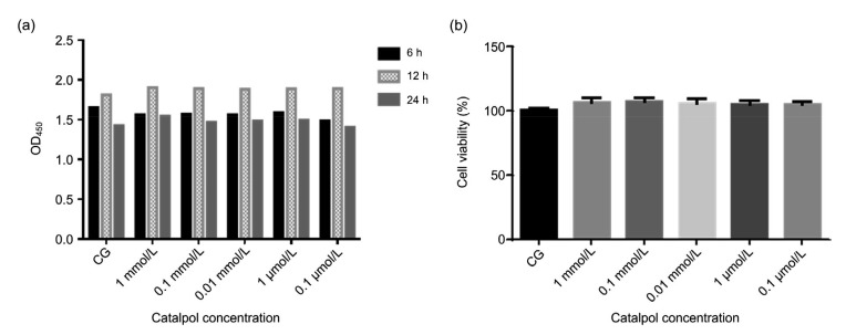
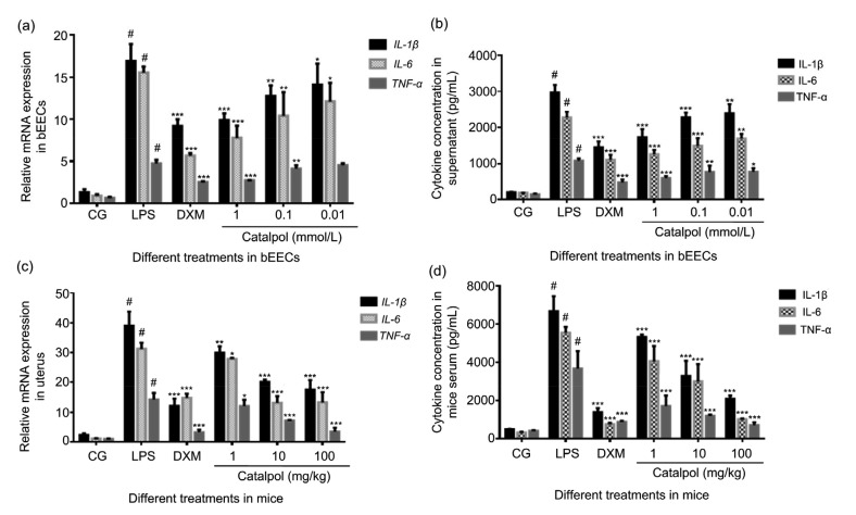
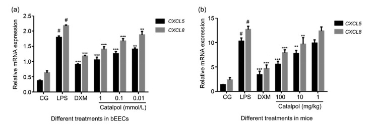
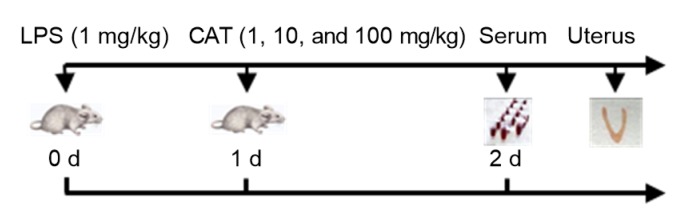
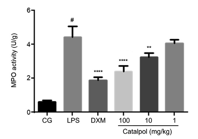
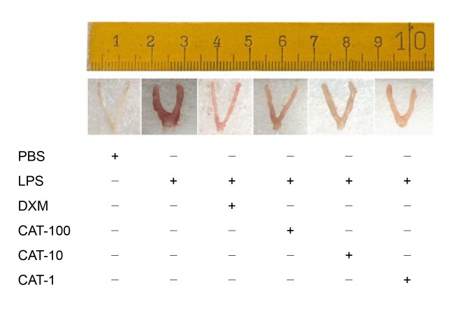
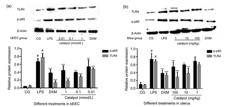
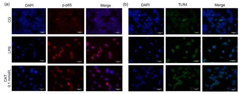
Catalpol is the main active ingredient of an extract from Radix rehmanniae, which in a previous study showed a protective effect against various types of tissue injury. However, a protective effect of catalpol on uterine inflammation has not been reported. In this study, to investigate the protective mechanism of catalpol on lipopolysaccharide (LPS)-induced bovine endometrial epithelial cells (bEECs) and mouse endometritis, in vitro and in vivo inflammation models were established. The Toll-like receptor 4 (TLR4)/nuclear factor-κB (NF-κB) signaling pathway and its downstream inflammatory factors were detected by enzyme-linked immunosorbent assay (ELISA), quantitative real-time polymerase chain reaction (qRT-PCR), western blot (WB), and immunofluorescence techniques. The results from ELISA and qRT-PCR showed that catalpol dose-dependently reduced the expression of pro-inflammatory cytokines such as tumor necrosis factor α (TNF-α), interleukin (IL)-1β, and IL-6, and chemokines such as C-X-C motif chemokine ligand 8 (CXCL8) and CXCL5, both in bEECs and in uterine tissue. From the experimental results of WB, qRT-PCR, and immunofluorescence, the expression of TLR4 and the phosphorylation of NF-κB p65 were markedly inhibited by catalpol compared with the LPS group. The inflammatory damage to the mouse uterus caused by LPS was greatly reduced and was accompanied by a decline in myeloperoxidase (MPO) activity. The results of this study suggest that catalpol can exert an anti-inflammatory impact on LPS-induced bEECs and mouse endometritis by inhibiting inflammation and activation of the TLR4/NF-κB signaling pathway.

Keywords: Catalpol; Endometritis; Inflammation; Toll-like receptor 4 (TLR4); Nuclear factor-κB (NF-κB).


Conflict of interest statement

Compliance with ethics guidelines: Hua ZHANG, Zhi-min WU, Ya-ping YANG, Aftab SHAUKAT, Jing YANG, Ying-fang GUO, Tao ZHANG, Xin-ying ZHU, Jin-xia QIU, Gan-zhen DENG, and Dong-mei SHI declare that they have no conflicts of interest.

All institutional and national guidelines for the care and use of laboratory animals were followed. The animal experiments were carried out according to the guidelines of the Laboratory Animal Research Center of Hubei Province and approved by the Ethical Committee on Animal Research at Huazhong Agricultural University (HZAUMO-2015-12), Wuhan, China.

Figures

Fig. 1
Fig. 1
Chemical structure of catalpol
Fig. 2
Fig. 2
Effect of catalpol on the viability of bEECs (a) Cells were cultured in 96-well cell plates at a density of 1×10⁵ cells/mL for 24 h, and then catalpol was added at five concentrations (1 mmol/L, 0.1 mmol/L, 0.01 mmol/L, 1 μmol/L, or 0.1 μmol/L), followed by incubation for 6, 12, and 24 h. After CCK-8 was added for 2 h, the optical density was measured at a wavelength of 450 nm (OD450). (b) Catalpol at five concentrations was added to bEEC cultures for 12 h. CCK-8 was then added and after 2 h, OD450 was measured. The results were calculated using the following formula: cell viability (%)=(ODtreatment–ODblank)/(ODcontrol–ODblank)×100%. Data represent the mean±standard error of mean (SEM) of triplicate experiments. CG: control group; bEEC: bovine endometrial epithelial cell; CCK-8: Cell Counting Kit-8
Fig. 3
Fig. 3
Effect of catalpol on cytokine expression in bEECs and mice stimulated by LPS (a, b) Cytokine expression in bEECs, stimulated by LPS (1 μg/mL) and co-treated with 1, 0.1, or 0.01 mmol/L catalpol in an incubator for 12 h, measured by qRT-PCR (a) and ELISA (b). DXM (1 μmol/L) was set as the positive control. (c, d) The mRNA expression and concentration of cytokines in mice uterine tissue and serum in the LPS (1 mg/kg) group, DXM (5 mg/kg) group, and three catalpol (1, 10, and 100 mg/kg) groups as above, were measured by qRT-PCR (c) and ELISA (d). Results are expressed as percentages relative to the untreated control after normalizing to GAPDH levels (n=3). Data represent the mean±standard error of mean (SEM) of triplicate experiments. # indicates P<0.05 vs. the control group. *, **, and *** indicate P<0.05, P<0.01, and P<0.001 vs. the LPS group, respectively. CG: control group; LPS: lipopolysaccharide; DXM: dexamethasone; bEEC: bovine endometrial epithelial cell
Fig. 4
Fig. 4
CXCL5 and CXCL8 gene expression in bEECs and uterine tissue challenged by LPS and co-treated with catalpol (a) CXCL5 and CXCL8 mRNA expression in bEECs treated by LPS alone and with catalpol at 1, 0.1, or 0.01 mmol/L for 12 h in cell culture, determined by qRT-PCR. DXM (1 μmol/L) was set as the positive control. (b) Mice were stimulated by LPS for 24 h, then injected intraperitoneally with 100, 10, or 1 mg/kg catalpol. Uterine tissues were collected 24 h later. DXM (5 mg/kg) was set as the positive control. CXCL5 and CXCL8 mRNA expression was determined by qRT-PCR. Results are expressed as percentages relative to the untreated control after normalizing to GAPDH levels (n=3). Data represent the mean±standard error of mean (SEM) of triplicate experiments. # indicates P<0.05 vs. CG. ** and *** indicate P<0.01 and P<0.001 vs. the LPS group, respectively. CXCL5: C-X-C motif chemokine ligand 5; CG: control group; LPS: lipopolysaccharide; DXM: dexamethasone; bEEC: bovine endometrial epithelial cell
Fig. 5
Fig. 5
Time axis of experimental animal treatment Catalpol was injected into mice at 1, 10, or 100 mg/kg, 1 d after intrauterine injection of 1 mg/kg LPS. The control group received an intrauterine injection of PBS. The serum and uterine tissues of mice were collected the next day. DXM (5 mg/kg) group was set as a positive control. LPS: lipopolysaccharide; CAT: catalpol; PBS: phosphate-buffered saline; DXM: dexamethasone
Fig. 6
Fig. 6
MPO activity in uterine tissue Mice groups were set as above. Uterine tissues were collected, homogenized in PBS, and centrifuged at 12 000 r/min for 15 min. The supernatants were measured according to the manufacturer’s instruction in the MPO activity kit. Data represent the mean±standard error of mean (SEM) of triplicate experiments. # indicates P<0.05 vs. CG. ** and **** indicate P<0.01 and P<0.0001 vs. the LPS group, respectively. CG: control group; LPS: lipopolysaccharide; DXM: dexamethasone; MPO: myeloperoxidase; PBS: phosphate-buffered saline
Fig. 7
Fig. 7
Effect of catalpol on endometrial inflammation of LPS-induced endometritis in mice Catalpol was injected into mice at 1, 10, or 100 mg/kg (CAT-1, CAT-10, or CAT-100) 1 d after intrauterine injection of 1 mg/kg LPS. The control group received an intrauterine injection of PBS. The DXM (5 mg/kg) group was set as a positive control. Uterine tissues of the mice were collected the next day, and immersed in 4% (0.04 g/mL) paraformaldehyde for H&E staining. PBS: phosphate-buffered saline; LPS: lipopolysaccharide; DXM: dexamethasone; CAT: catalpol; H&E: hematoxylin and eosin
Fig. 8
Fig. 8
H&E staining of uterine tissue in control, LPS-induced and catalpol (10 mg/kg) co-treated mice (a, b) Control group. Histopathology of the endometrium from healthy mice. Black arrows point to the intact epithelium layer and red arrows to vacuoles. (c, d) LPS group. Histopathology of the endometrium from LPS-induced mice. Pathological changes are shown as hemorrhage in multiple areas (blue arrows), epithelial cell exfoliation (purple arrows), and a large amount of inflammatory cell aggregation (green arrows). The epithelial layer was severely damaged (yellow arrows). (e, f) LPS+catalpol group. Histopathology of the endometrium from LPS-induced mice co-treated with catalpol. Only a small part of the luminal epithelium was damaged (white arrows). The morphology of epithelial cells changed greatly, most becoming cubic in shape (brown arrows). A few inflammatory cells were seen in the epithelial layer (green arrows). LPS: lipopolysaccharide; H&E: hematoxylin and eosin
Fig. 9
Fig. 9
Effects of catalpol on the expression of TLR4 and p-p65 proteins in LPS-induced bEECs and mice uterine tissues (a) bEECs were pretreated with catalpol at 1, 0.1, or 0.01 mmol/L for 1 h, and then LPS (1 µg/mL) was added, followed by incubation for a further 12 h. DXM (1 μmol/L) group was set as a positive control. (b) Catalpol was injected into mice at 1, 10, or 100 mg/kg, 1 d after intrauterine injection of 1 mg/kg LPS. The control group received an intrauterine injection of PBS. DXM (5 mg/kg) group was set as a positive control. Uterine tissues of mice were collected the next day. The phosphorylation levels of p65 and TLR4 were analyzed by western blot. Immunoblot signals were detected by an enhanced chemiluminescence detection system. Quantification of protein expression was normalized to β-actin using a densitometer (n=3). The data represent the mean±standard error of mean (SEM) of triplicate experiments. # indicates P<0.05 vs. CG. ** and *** indicate P<0.01 and P<0.001 vs. the LPS group, respectively. CG: control group; LPS: lipopolysaccharide; DXM: dexamethasone; PBS: phosphate-buffered saline; bEEC: bovine endometrial epithelial cell; TLR4: Toll-like receptor 4
Fig. 10
Fig. 10
Immunofluorescence detection of p-p65 and TLR4 in bEECs The protein expression of p-p65 (a) and TLR4 (b) was detected by cell immunofluorescence assay in the control, LPS, and catalpol (0.1 mmol/L) groups. Bar=50 µm. CG: control group; LPS: lipopolysaccharide; CAT: catalpol; bEEC: bovine endometrial epithelial cell; DAPI: 4',6-diamidino-2-phenylindole; TLR4: Toll-like receptor 4

References

    1. Angrisano T, Pero R, Peluso S, et al. LPS-induced IL-8 activation in human intestinal epithelial cells is accompanied by specific histone H3 acetylation and methylation changes. BMC Microbiol, 10:172. 2010 doi: 10.1186/1471-2180-10-172. - DOI - PMC - PubMed
    1. Begley LA, Kasina S, Mehra R, et al. CXCL5 promotes prostate cancer progression. Neoplasia. 2008;10(3):244–254. doi: 10.1593/neo.07976. - DOI - PMC - PubMed
    1. Brodzki P, Kostro K, Brodzki A, et al. Inflammatory cytokines and acute-phase proteins concentrations in the peripheral blood and uterus of cows that developed endometritis during early postpartum. Theriogenology. 2015;84(1):11–18. doi: 10.1016/j.theriogenology.2015.02.006. - DOI - PubMed
    1. Brown K, Park S, Kanno T, et al. Mutual regulation of the transcriptional activator NF-κB and its inhibitor, IκBα. Proc Natl Acad Sci USA. 1993;90(6):2532–2536. doi: 10.1073/pnas.90.6.2532. - DOI - PMC - PubMed
    1. Chen BY, Jiang LX, Hao K, et al. Protection of plasma transfusion against lipopolysaccharide/D-galactosamine-induced fulminant hepatic failure through inhibiting apoptosis of hepatic cells in mice. J Zhejiang Univ-Sci B (Biomed & Biotechnol) 2018;19(6):436–444. doi: 10.1631/jzus.B1700277. - DOI