. 2019 Sep 6;20(18):4375.
doi: 10.3390/ijms20184375.

Gentamicin Targets Acid Sphingomyelinase in Cancer: The Case of the Human Gastric Cancer NCI-N87 Cells

Affiliations

Gentamicin Targets Acid Sphingomyelinase in Cancer: The Case of the Human Gastric Cancer NCI-N87 Cells

Elisabetta Albi et al. Int J Mol Sci. .

Abstract

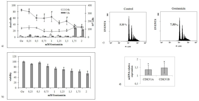
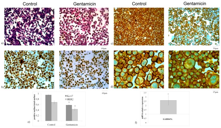
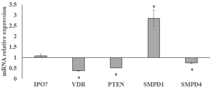
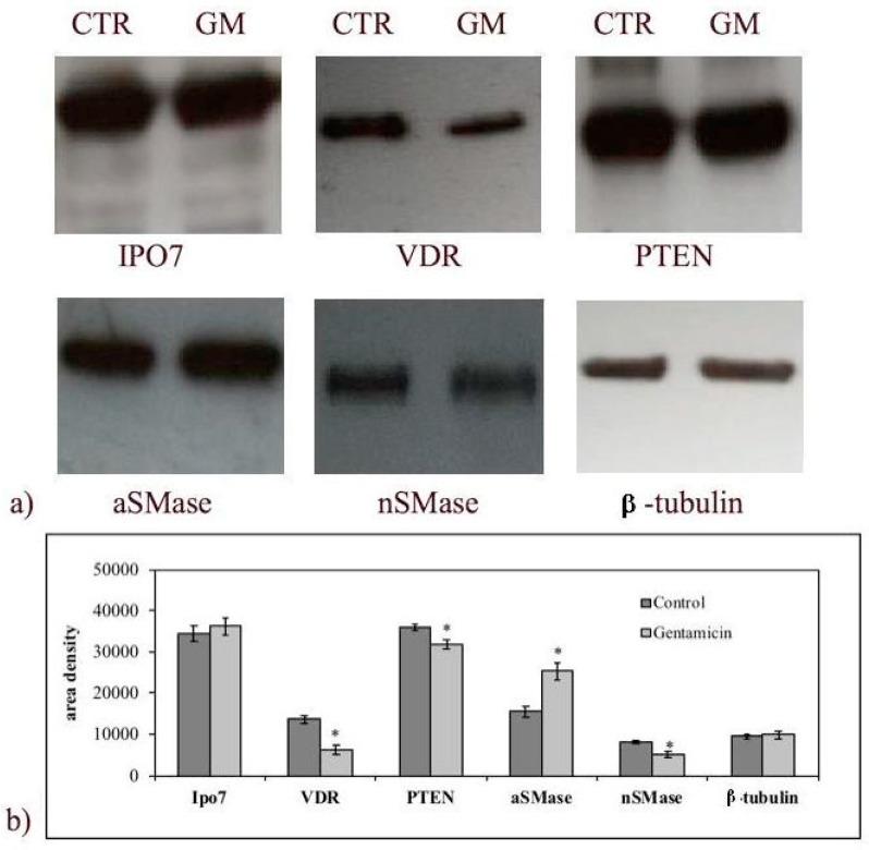
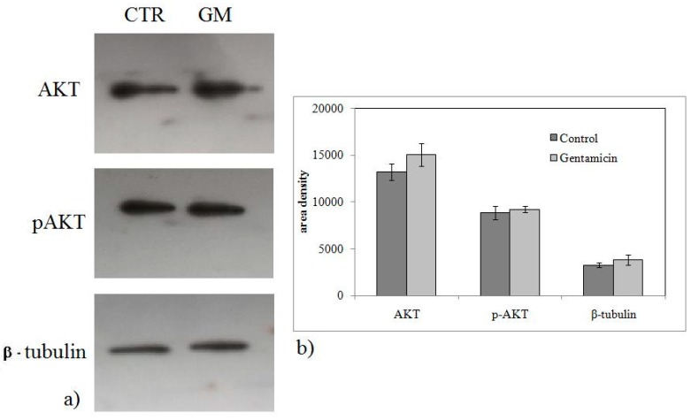
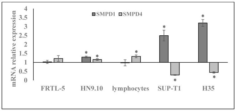
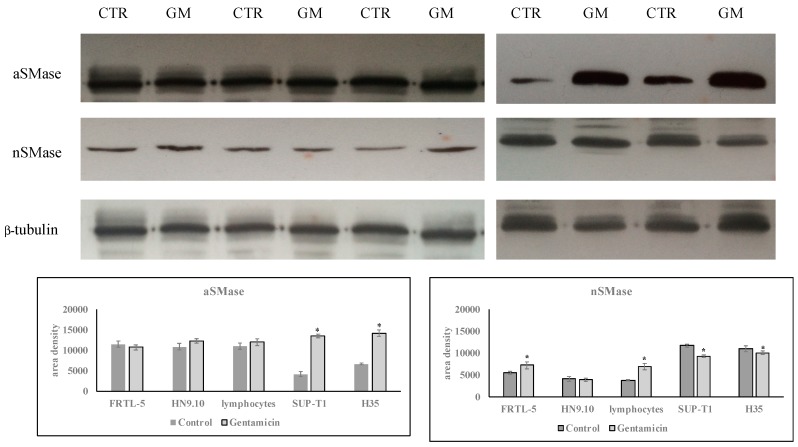
Emerging literature implicates acid sphingomyelinase in tumor sensitivity/resistance to anticancer treatments. Gentamicin is a drug commonly used as an antimicrobial but its serendipity effects have been shown. Even though many evidences on the role of gentamicin in cancer have been reported, its mechanism of action is poorly understood. Here, we explored acid sphingomyelinase as a possible new target of gentamicin in cancer. Since gastric cancer is one of the most common cancers and represents the second cause of death in the world, we performed the study in NCI-N87 gastric cancer cell line. The effect of the drug resulted in the inhibition of cell proliferation, including a reduction of cell number and viability, in the decrease of MIB-1 proliferative index as well as in the upregulation of cyclin-dependent kinase inhibitor 1A and 1B (CDKN1A and CDKN1B), and growth arrest and DNA-damage 45A (GADD45A) genes. The cytotoxicity was apoptotic as shown by FACS analysis. Additionally, gentamicin reduced HER2 protein, indicating a minor tumor aggressiveness. To further define the involvement of sphingomyelin metabolism in the response to the drug, gene and protein expression of acid and neutral sphingomeylinase was analyzed in comparison with phosphatase and tensin homolog deleted on chromosome 10 (PTEN) and vitamin D receptor (VDR), molecules involved in cancer. Gentamicin induced a downregulation of PTEN, VDR, and neutral sphingomyelinase and a strong upregulation of acid sphingomyelinase. Of note, we identified the same upregulation of acid sphingomyelinase upon gentamicin treatment in other cancer cells and not in normal cells. These findings provide new insights into acid sphingomyelinase as therapeutic target, reinforcing studies on the potential role of gentamicin in anticancer therapy.

Keywords: cancer; drug target; lipid; sphingomyelin; sphingomyelinase.


Conflict of interest statement

The authors declare no conflict of interest.

Figures

Figure 1
Figure 1
Effects of increasing doses of gentamicin on NCI-N87 cells. (a) The cells were counted 24 and 72 h after treatment with increasing concentrations of the drug (0.25–2.0 mM) (lines referred to the ordinate on the right). Cell death was evaluated by trypan blue staining (histograms referred to the ordinate on the left). Ctr, untreated sample. (b) Cell viability was assessed by MTT method and expressed as a percentage relative to that of the control cells set at 100%. (c) FACS analysis. (d) CDKN1A, CDKN1B gene expression, data are referred to the gentamicin (GM)-untreated sample (control) set at 1. GAPDH expression was used as a housekeeping gene. RT-PCR analysis was performed in control and experimental NCI-N87 cells collected 24 h after 1.5 mM GM treatment. The data represent the mean ± standard deviation (SD) of three independent experiments performed in duplicate (significance, * p < 0.001 versus control sample).
Figure 2
Figure 2
Effect of gentamicin after 24 h from treatment. (a) Cell morphology by hematoxylin–eosin staining; (b) MIB-1 immunohistochemistry staining; (c,d) HercepTest immunostaining. 10× magnification (a,b), 20× magnification (c), and 40× magnification (d); (e) densitometric analysis; (f) GADD45A gene expression, data are referred to the GM-untreated sample (control) set at 1. GAPDH expression was used as a housekeeping gene. RT-PCR analysis was performed in control and experimental NCI-N87 cells collected 24 h after 1.5 mM GM treatment. The data represent the mean ± SD of three independent experiments performed in duplicate (significance, * p < 0.001 versus control sample).
Figure 3
Figure 3
Effect of gentamicin on Ipo7, VDR, PTEN, SMPD1, SMPD4, gene expression. GAPDH expression was used as a housekeeping gene. RT-PCR analysis was performed in control and experimental NCI-N87 cells collected 24 h after 1.5 mM GM treatment. Data are expressed as the mean ± SD of three independent experiments performed in three PCR replicates (significance, * p < 0.001 versus control sample).
Figure 4
Figure 4
Effect of gentamicin on importin 7 (Ipo7), vitamin D receptor (VDR), phosphatase and tensin homolog deleted on chromosome 10 (PTEN), acid sphingomyelinase (aSMase), and neutral sphingomyelinase (nSMase) protein expression. Experiments were performed in control and experimental NCI-N87 cells collected 24 h after 1.5 mM GM treatment. (a) Immunoblots of proteins were probed with anti-IPO7, anti-VDR, anti-PTEN, anti-aSMase, and anti-nSMase and visualized by enhanced chemiluminescence (ECL). β-tubulin was used as loading control. (b) The area intensity was evaluated by densitometry scanning and analysis with ImageJ program, the data represent the mean ± SD of three experiments performed in duplicate (significance, * p < 0.001 versus control sample).
Figure 5
Figure 5
Effect of gentamicin on AKT/p-AKP protein expression. Experiments were performed in control and experimental NCI-N87 cells collected 24 h after 1.5 mM GM treatment. (a) Immunoblots of proteins were probed with anti-AKT and p-AKT and visualized by enhanced chemiluminescence (ECL). β-tubulin was used as loading control. (b) The area density was evaluated by densitometry scanning and analysis with ImageJ program, the data represent the mean ± SD of three experiments performed in duplicate (significance, * p < 0.001 versus control sample).
Figure 6
Figure 6
Effect of gentamicin on SMPD1, SMPD4 gene expression in thyrocytes (FRTL-5 cells), embryonic hippocampal cells (HN9.10), human lymphocytes and non-Hodgkin’s T cell human lymphoblastic lymphoma cells (SUP-T1), hepatoma cells (H35), and human gastric cancer cells (NCI-N87). GAPDH expression was used as a housekeeping gene. RT-PCR analysis was performed in control and experimental cells collected after 24 h after 1.5mM GM treatment. Data are expressed as the mean ± SD of three independent experiments performed in three PCR replicates (significance, * p < 0.001 versus control sample).
Figure 7
Figure 7
Effect of gentamicin on aSMase and nSMase protein expression. Experiments were performed in control and experimental FRTL-5, HN9.10, lymphocytes, SUP-T1, and H35 cells, collected 24 h after 1.5 mM GM treatment. (a) Immunoblots of proteins were probed with anti-aSMase and anti-nSMase and visualized by enhanced chemiluminescence (ECL). β-tubulin was used as loading control. (b) The area intensity was evaluated by densitometry scanning and analysis with ImageJ program, the data represent the mean ± SD of three experiments performed in duplicate (significance, * p < 0.001 versus control sample).

References

    1. Testi R. Sphingomyelin breakdown and cell fate. Trends Biochem. Sci. 1996;21:468–471. doi: 10.1016/S0968-0004(96)10056-6. - DOI - PubMed
    1. Albi E. Role of intranuclear lipids in health and disease. Clin. Lipidol. 2011;6:59–69. doi: 10.2217/clp.10.83. - DOI
    1. Canals D., Perry D.M., Jenkins R.W., Hannun Y.A. Drug targeting of sphingolipid metabolism: Sphingomyelinases and ceramidases. Br. J. Pharmacol. 2011;163:694–712. doi: 10.1111/j.1476-5381.2011.01279.x. - DOI - PMC - PubMed
    1. Marchesini N., Luberto C., Hannun Y.A. Biochemical properties of mammalian neutral sphingomyelinase 2 and its role in sphingolipid metabolism. J. Biol. Chem. 2003;278:13775–13783. doi: 10.1074/jbc.M212262200. - DOI - PubMed
    1. Ito H., Murakami M., Furuhata A., Gao S., Yoshida K., Sobue S., Hagiwara K., Takagi A., Kojima T., Suzuki M., et al. Transcriptional regulation of neutral sphingomyelinase 2 gene expression of a human breast cancer cell line, MCF-7, induced by the anti-cancer drug, daunorubicin. Biochim. Biophys. Acta. 2009;1789:681–690. doi: 10.1016/j.bbagrm.2009.08.006. - DOI - PubMed

MeSH terms