. 2019 Sep 6;11(9):829.
doi: 10.3390/v11090829.

No Evidence for a Role for Antibodies during Vaccination-Induced Enhancement of Porcine Reproductive and Respiratory Syndrome

Affiliations

No Evidence for a Role for Antibodies during Vaccination-Induced Enhancement of Porcine Reproductive and Respiratory Syndrome

Carmen A Sautter et al. Viruses. .

Abstract

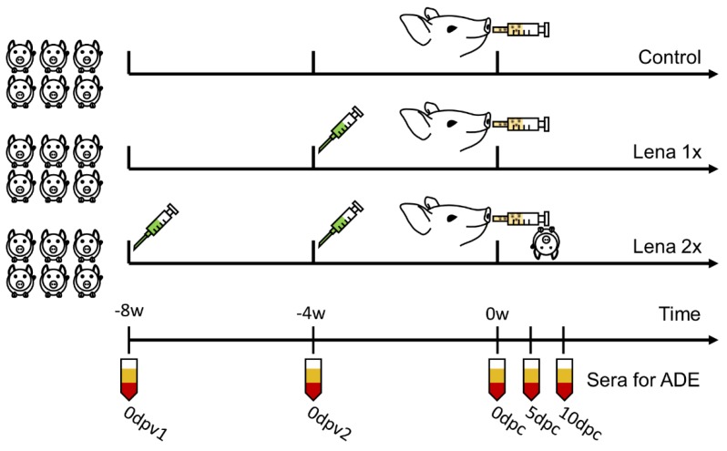
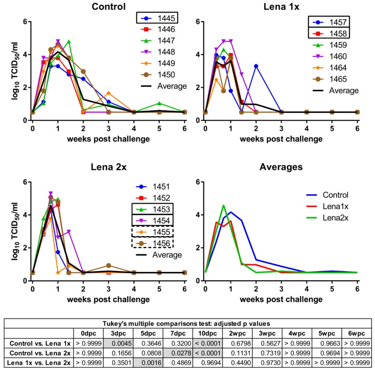
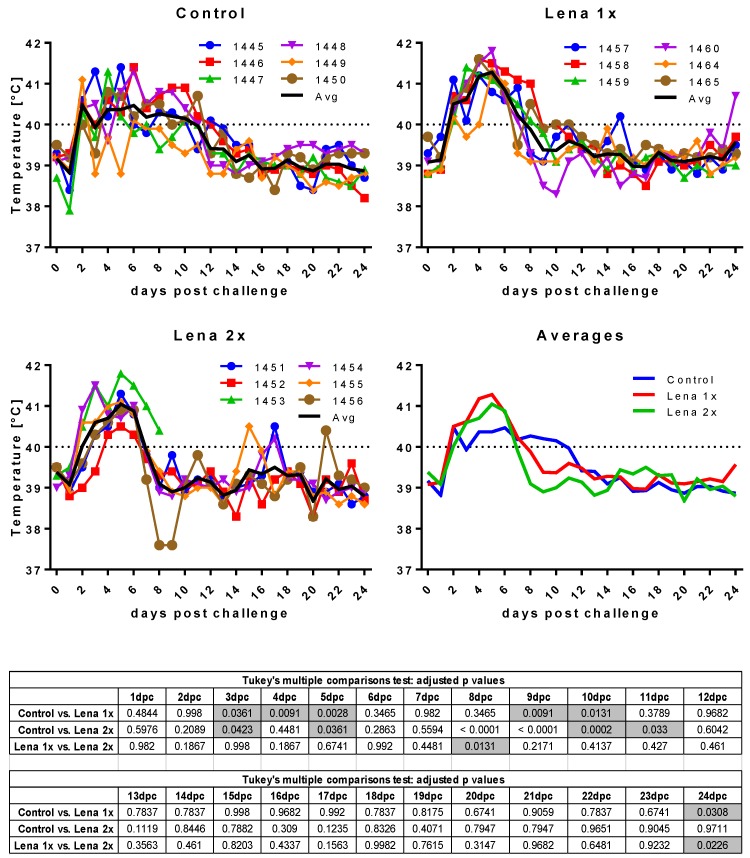
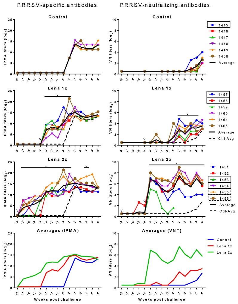
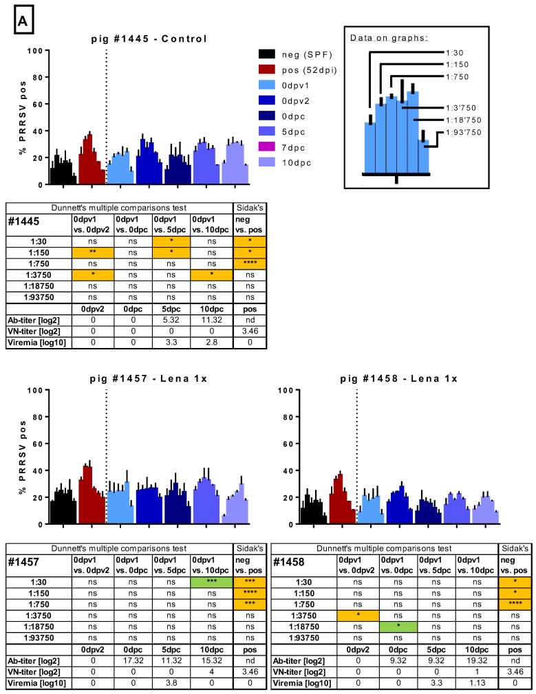
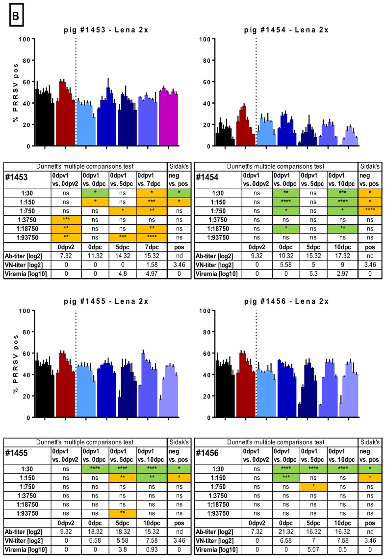
Vaccination is one of the most important tools to protect pigs against infection with porcine reproductive and respiratory syndrome virus 1 (PRRSV-1). Although neutralizing antibodies are considered to represent an important mechanism of protective immunity, anti-PRRSV antibodies, in particular at subneutralizing concentrations, have also been reported to exacerbate PRRSV infection, probably through FcγR-mediated uptake of antibody-opsonized PRRSV, resulting in enhanced infection of, and replication in, target cells. Therefore, we investigated this pathway using sera from an animal experiment in which vaccine-mediated enhancement of clinical symptoms was observed. Three groups of six pigs were vaccinated with an inactivated PRRSV vaccine based on the PRRSV-1 subtype 3 strain Lena and challenged after a single or a prime-boost immunization protocol, or injected with PBS. We specifically tested if sera obtained from these animals can enhance macrophage infections, viral shedding, or cytokine release at different dilutions. Neither the presence of neutralizing antibodies nor general anti-PRRSV antibodies, mediated an enhanced infection, increased viral release or cytokine production by macrophages. Taken together, our data indicate that the exacerbated disease was not caused by antibodies.

Keywords: PRRSV-1; antibody-dependent enhancement (ADE) of disease; homologous challenge; in vivo; inactivated vaccine; monocyte-derived macrophages.


Conflict of interest statement

The authors declare no conflict of interest. The funders had no role in the design of the study; in the collection, analyses, or interpretation of data; in the writing of the manuscript, or in the decision to publish the results.

Figures

Figure A1
Figure A1
Experiment layout of vaccination and challenge.
Figure 1
Figure 1
Viremia of Lena-vaccinated pigs following homologous challenge infection. Viremia was determined by titration on PAMs collected from porcine reproductive and respiratory syndrome virus (PRRSV)-negative pigs and IPMA. Pigs in the control group received two injections of PBS + adjuvant, eight and four weeks before the challenge. Group “Lena 1x” was vaccinated once with inactivated Lena, four weeks before the challenge. The group “Lena 2x” was vaccinated twice at eight and four weeks before the challenge. dpc—days post-challenge, wpc—weeks post-challenge. Numbers (1451–1465) represent the individual pigs. Solid rectangles around pig numbers mean they have been used in all ADE experiments, dashed rectangles mean that their sera have also been further analyzed, but not in all ADE experiments. The black line is the average of each group. Statistical analysis was performed with RM 2-way ANOVA, followed by Tukey’s multiple comparison test. Statistically significant values are highlighted in grey in the table under the graphs.
Figure 2
Figure 2
Body temperature after challenge. Control, Lena 1x, and Lena 2x: Colored lines represent individual animals and thick black lines represent the mean value for each group. Temperature >40 °C was considered as fever (dotted line on upper panel). dpc—days post-challenge. Statistical analysis was performed with an ordinary 2-way ANOVA, followed by Tukey’s multiple comparison test. Statistically significant values are highlighted in grey in the table under the graphs.
Figure 3
Figure 3
Respiratory disease scores after challenge. Control, Lena 1x, and Lena 2x: Colored lines represent individual animals and thick black lines represent the mean value for each group. Scores: 0 = normal; 1 = mild dyspnea and/or tachypnea when stressed; 2 = mild dyspnea and/or tachypnea at rest; 3 = moderate dyspnea and/or tachypnea when stressed; 4 = moderate dyspnea and/or tachypnea at rest; 5 = severe dyspnea and/or tachypnea when stressed; 6 = severe dyspnea and/or tachypnea at rest. dpc—days post-challenge. Statistical analysis was performed with an ordinary 2-way ANOVA, followed by Tukey’s multiple comparison test. Statistically significant values are highlighted in grey in the table under the graphs.
Figure 4
Figure 4
PRRSV-specific IPMA (left) and VN (right) antibodies before and after challenge 105 TCID50 PRRSV Lena. Control pigs received two injections of PBS in adjuvant, at eight and four weeks before the challenge. Group “Lena 1x” was vaccinated once, four weeks before the challenge. Group “Lena 2x” received a prime/booster vaccination at eight and four weeks before the challenge. PRRSV-specific antibodies were detected by IPMA. PRRSV-neutralizing antibodies were detected by a virus neutralization assay (VNT). Colored lines with symbols represent individual pigs. Solid black lines represent the average of each group and dashed black lines represent the average of the control group. v—vaccination; numbers represent individual pig numbers. Solid rectangles around pig numbers mean they have been used in all ADE experiments, dashed rectangles mean their sera have also been further analyzed, but not in all ADE experiments. Statistical analysis was performed with RM 2-way ANOVA, followed by Tukey’s multiple comparison test. The comparison was: Lena 1x vs. Control, and Lena 2x vs. Control. p < 0.05 was considered statistically significant and is represented by *.
Figure 5
Figure 5
Macrophage infection in the presence of immune sera. Sera of several randomly selected pigs from the different groups were tested for their ability to enhance the infection of monocyte-derived macrophages (MDMs). Serial dilutions (1:30 to 1:93,750) of the sera were pre-incubated with Lena. Then, MDMs were infected in triplicates and tested PRRSV N-protein after 16 h by flow cytometry. In A, the results for pig #1445, from the control group, which received 2 injections of PBS + adjuvant, as well as the results for pigs #1457 and #1458, from the “Lena 1x” group, which were vaccinated once, is shown. In B. the results for pigs #1453–1456, from the “Lena 2x” group, which were vaccinated twice, is shown. Bars show mean with SD. dpv—days post-vaccination, dpc—days post-challenge, dpi—days post-infection; 0dpv1 = eight weeks before challenge, 0dpv2 = four weeks before challenge. Statistical analysis was performed with ordinary 2-way ANOVA, followed by Dunnett’s multiple comparison test or Sidak’s post hoc test, as indicated. Green highlighting indicates a neutralizing effect, while orange highlighting indicates an enhancing effect. Symbols: * = p < 0.05, ** = p < 0.01, *** = p < 0.001, **** = p < 0.0001.
Figure 5
Figure 5
Macrophage infection in the presence of immune sera. Sera of several randomly selected pigs from the different groups were tested for their ability to enhance the infection of monocyte-derived macrophages (MDMs). Serial dilutions (1:30 to 1:93,750) of the sera were pre-incubated with Lena. Then, MDMs were infected in triplicates and tested PRRSV N-protein after 16 h by flow cytometry. In A, the results for pig #1445, from the control group, which received 2 injections of PBS + adjuvant, as well as the results for pigs #1457 and #1458, from the “Lena 1x” group, which were vaccinated once, is shown. In B. the results for pigs #1453–1456, from the “Lena 2x” group, which were vaccinated twice, is shown. Bars show mean with SD. dpv—days post-vaccination, dpc—days post-challenge, dpi—days post-infection; 0dpv1 = eight weeks before challenge, 0dpv2 = four weeks before challenge. Statistical analysis was performed with ordinary 2-way ANOVA, followed by Dunnett’s multiple comparison test or Sidak’s post hoc test, as indicated. Green highlighting indicates a neutralizing effect, while orange highlighting indicates an enhancing effect. Symbols: * = p < 0.05, ** = p < 0.01, *** = p < 0.001, **** = p < 0.0001.
Figure 6
Figure 6
Impact of sera on virus titers in MDM cultures. Serially diluted immune sera were pre-incubated with Lena. Then, MDMs were infected with these immune complex mixtures, washed, and incubated for a total of 16 h. The supernatants were titrated in triplicates. Controls 1–3 show the viral titers when PRRSV-Ab-negative serum (neg) or PRRSV-immune serum (pos, from 52 dpi) were used as immune sera. Pig #1445, from the control group, had not been immunized before challenge. Pigs #1457 and #1458, from the “Lena 1x” group, were vaccinated once with BEI-inactivated Lena + adjuvant, and pig #1454, from the “Lena 2x” group, was vaccinated twice with the same vaccine. dpv—days post-vaccination, dpc—days post-challenge, dpi—days post-infection; 0dpv1 = eight weeks before challenge, 0dpv2 = four weeks before challenge. Statistical analysis was performed with ordinary 2-way ANOVA, followed by Tukey’s multiple comparison test. Within each dilution, the sera from different time points were compared with the “initial serum” (neg or 0dpv1). Symbols: * = p < 0.05, ** = p < 0.01, *** = p < 0.001, **** = p < 0.0001. Color of the asterisks denote to which serum they belong.

Similar articles

Cited by

References

    1. Cafruny W.A., Plagemann P.G. Immune response to lactate dehydrogenase-elevating virus: Serologically specific rabbit neutralizing antibody to the virus. Infect. Immun. 1982;37:1007–1012. - PMC - PubMed
    1. Takeda A., Tuazon C.U., Ennis F.A. Antibody-enhanced infection by hiv-1 via fc receptor-mediated entry. Science. 1988;242:580–583. doi: 10.1126/science.2972065. - DOI - PubMed
    1. Homsy J., Meyer M., Tateno M., Clarkson S., Levy J.A. The fc and not cd4 receptor mediates antibody enhancement of hiv infection in human cells. Science. 1989;244:1357–1360. doi: 10.1126/science.2786647. - DOI - PubMed
    1. Halstead S.B., Porterfield J.S., O’Rourke E.J. Enhancement of dengue virus infection in monocytes by flavivirus antisera. Am. J. Trop. Med. Hyg. 1980;29:638–642. doi: 10.4269/ajtmh.1980.29.638. - DOI - PubMed
    1. Halstead S.B. Dengue antibody-dependent enhancement: Knowns and unknowns. Microbiol. Spectr. 2014;2 doi: 10.1128/microbiolspec. - DOI - PubMed

Publication types

MeSH terms