

Flow Cytometry Reveals the Nature of Oncotic Cells
- PMID: 31489916
- PMCID: PMC6769836
- DOI: 10.3390/ijms20184379
Flow Cytometry Reveals the Nature of Oncotic Cells
Abstract
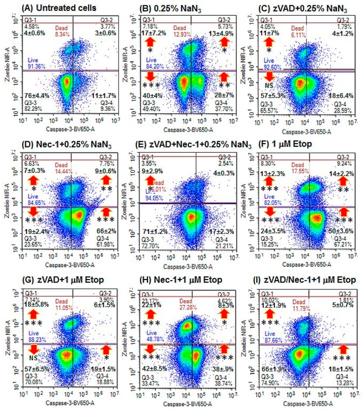
The term necrosis is commonly applied to cells that have died via a non-specific pathway or mechanism but strictly is the description of the degradation processes involved once the plasma membrane of the cell has lost integrity. The signalling pathways potentially involved in accidental cell death (ACD) or oncosis are under-studied. In this study, the flow cytometric analysis of the intracellular antigens involved in regulated cell death (RCD) revealed the phenotypic nature of cells undergoing oncosis or necrosis. Sodium azide induced oncosis but also classic apoptosis, which was blocked by zVAD (z-Vla-Ala-Asp(OMe)-fluoromethylketone). Oncotic cells were found to be viability+ve/caspase-3-ve/RIP3+ve/-ve (Receptor-interacting serine/threonine protein kinase 3). These two cell populations also displayed a DNA damage response (DDR) phenotype pH2AX+ve/PARP-ve, cleaved PARP induced caspase independent apoptosis H2AX-ve/PARP+ve and hyper-activation or parthanatos H2AX+ve/PARP+ve. Oncotic cells with phenotype cell viability+ve/RIP3-ve/caspase-3-ve showed increased DDR and parthanatos. Necrostatin-1 down-regulated DDR in oncotic cells and increased sodium azide induced apoptosis. This flow cytometric approach to cell death research highlights the link between ACD and the RCD processes of programmed apoptosis and necrosis.
Keywords: DDR; accidental cell death; flow cytometry; oncosis; parthanatos.
Conflict of interest statement
A.V. received salary funding from GSK. GSK had no input or role in the conception or undertaking of this study in any manner. Other authors declare no conflict of interest. The funders had no role in the design of the study; in the collection, analyses, or interpretation of data; in the writing of the manuscript, or in the decision to publish the results.
Figures


Similar articles
-
Simultaneous polychromatic flow cytometric detection of multiple forms of regulated cell death.Apoptosis. 2019 Jun;24(5-6):453-464. doi: 10.1007/s10495-019-01528-w. Apoptosis. 2019. PMID: 30788651 Free PMC article.
-
Flow cytometric detection of hyper-polarized mitochondria in regulated and accidental cell death processes.Apoptosis. 2020 Aug;25(7-8):548-557. doi: 10.1007/s10495-020-01613-5. Apoptosis. 2020. PMID: 32495124 Free PMC article.
-
Simultaneous flow cytometric immunophenotyping of necroptosis, apoptosis and RIP1-dependent apoptosis.Methods. 2018 Feb 1;134-135:56-66. doi: 10.1016/j.ymeth.2017.10.013. Epub 2017 Nov 24. Methods. 2018. PMID: 29175336
-
Multiparametric flow cytometric analysis of biochemical and functional events associated with apoptosis and oncosis using the 7-aminoactinomycin D assay.J Immunol Methods. 2002 Jul 1;265(1-2):81-96. doi: 10.1016/s0022-1759(02)00072-8. J Immunol Methods. 2002. PMID: 12072180 Review.
-
Unravelling oncosis: morphological and molecular insights into a unique cell death pathway.Front Immunol. 2024 Aug 29;15:1450998. doi: 10.3389/fimmu.2024.1450998. eCollection 2024. Front Immunol. 2024. PMID: 39281670 Free PMC article. Review.
Cited by
-
Ferroptosis as a novel form of regulated cell death: Implications in the pathogenesis, oncometabolism and treatment of human cancer.Genes Dis. 2020 Dec 4;9(2):347-357. doi: 10.1016/j.gendis.2020.11.019. eCollection 2022 Mar. Genes Dis. 2020. PMID: 35224151 Free PMC article. Review.
-
Application of Regulatory Cell Death in Cancer: Based on Targeted Therapy and Immunotherapy.Front Immunol. 2022 Mar 10;13:837293. doi: 10.3389/fimmu.2022.837293. eCollection 2022. Front Immunol. 2022. PMID: 35359956 Free PMC article. Review.
-
The Role of Death-Associated Protein Kinase-1 in Cell Homeostasis-Related Processes.Genes (Basel). 2023 Jun 16;14(6):1274. doi: 10.3390/genes14061274. Genes (Basel). 2023. PMID: 37372454 Free PMC article. Review.
-
Novel 2-(5-Arylthiophen-2-yl)-benzoazole Cyclometalated Iridium(III) dppz Complexes Exhibit Selective Phototoxicity in Cancer Cells by Lysosomal Damage and Oncosis.J Med Chem. 2024 Jan 11;67(1):691-708. doi: 10.1021/acs.jmedchem.3c01978. Epub 2023 Dec 23. J Med Chem. 2024. PMID: 38141031 Free PMC article.
References
-
- Galluzzi L., Bravo-San Pedro J.M., Vitale I., Aaronson S.A., Abrams J.M., Adam D., Alnemri E.S., Altucci L., Andrews D., Annicchiarico-Petruzzelli M., et al. Essential versus accessory aspects of cell death: Recommendations of the NCCD 2015. Cell Death Differ. 2015;22:58–73. doi: 10.1038/cdd.2014.137. - DOI - PMC - PubMed
-
- Galluzzi L., Vitale I., Aaronson S.A., Abrams J.M., Adam D., Agostinis P., Alnemri E.S., Altucci L., Amelio I., Andrews D.W., et al. Molecular mechanisms of cell death: Recommendations of the Nomenclature Committee on Cell Death 2018. Cell Death Differ. 2018;25:486–541. doi: 10.1038/s41418-017-0012-4. - DOI - PMC - PubMed
-
- Morales J., Li L., Fattah F.J., Dong Y., Bey E.A., Patel M., Gao J., Boothman D.A. Review of Poly (ADP-ribose) Polymerase (PARP) Mechanisms of Action and Rationale for Targeting in Cancer and Other Diseases. Crit. Rev. Eukaryot. Gene Expr. 2014;24:15–28. doi: 10.1615/CritRevEukaryotGeneExpr.2013006875. - DOI - PMC - PubMed
MeSH terms
Substances
LinkOut - more resources
Full Text Sources
Research Materials
Miscellaneous
