. 2019 Sep 6;14(9):e0214829.
doi: 10.1371/journal.pone.0214829. eCollection 2019.

Tamoxifen suppresses pancreatic β-cell proliferation in mice

Affiliations

Tamoxifen suppresses pancreatic β-cell proliferation in mice

Surl-Hee Ahn et al. PLoS One. .

Abstract

Tamoxifen is a mixed agonist/antagonist estrogen analogue that is frequently used to induce conditional gene deletion in mice using Cre-loxP mediated gene recombination. Tamoxifen is routinely employed in extremely high-doses relative to typical human doses to induce efficient gene deletion in mice. Although tamoxifen has been widely assumed to have no influence upon β-cells, the acute developmental and functional consequences of high-dose tamoxifen upon glucose homeostasis and adult β-cells are largely unknown. We tested if tamoxifen influences glucose homeostasis in male mice of various genetic backgrounds. We then carried out detailed histomorphometry studies of mouse pancreata. We also performed gene expression studies with islets of tamoxifen-treated mice and controls. Tamoxifen had modest effects upon glucose homeostasis of mixed genetic background (F1 B6129SF1/J) mice, with fasting hyperglycemia and improved glucose tolerance but without overt effects on fed glucose levels or insulin sensitivity. Tamoxifen inhibited proliferation of β-cells in a dose-dependent manner, with dramatic reductions in β-cell turnover at the highest dose (decreased by 66%). In sharp contrast, tamoxifen did not reduce proliferation of pancreatic acinar cells. β-cell proliferation was unchanged by tamoxifen in 129S2 mice but was reduced in C57Bl6 genetic background mice (decreased by 59%). Gene expression studies revealed suppression of RNA for cyclins D1 and D2 within islets of tamoxifen-treated mice. Tamoxifen has a cytostatic effect on β-cells, independent of changes in glucose homeostasis, in mixed genetic background and also in C57Bl6 mice. Tamoxifen should be used judiciously to inducibly inactivate genes in studies of glucose homeostasis.


Conflict of interest statement

JAK currently serves as medical director of McNair Interests, a private equity group with investments in type 1 diabetes and other chronic illnesses and is also an advisor for Sanofi and Lexicon. CJL declares no conflict of interest relevant to this article. MMR currently serves as a full time employee of Janssen Research and Development, Johnson and Johnson, Springhouse, PA. AG currently serves as a full time employee of the Novartis Institutes for BioMedical Research (NIBR), a subsidiary of Novartis International AG, Cambridge, MA. No other potential conflicts of interest relevant to this article were reported. This does not alter our adherence to PLOS ONE policies on sharing data and materials.

Figures

Fig 1
Fig 1. Tamoxifen increases fasting glucose but improves glucose tolerance in mixed genetic background (F1 hybrid B6129SF1/J) mice.
(a) Schematic indicating time course of treatment. Mice received daily oral gavage with tamoxifen (0.025, 0.1, 0.2, or 0.3mg/g body weight) or vehicle for 5 days. Mice were sequentially labeled with BrdU for two weeks each, starting immediately after tamoxifen treatment. (b, c) Body weight (b) and blood glucose (c) change from the start of treatment (day -5) to the end of the experiment (day 14). Random fed (d) and fasting (e) blood glucose levels at the end of the experiment. (f, g) Glucose tolerance tests (GTT) 5 days after the end of tamoxifen with glucose curves (f) and GTT area under curve (AUC) calculations (g). (h, i) Insulin tolerance tests (ITT) 14 days after tamoxifen with glucose curves (h) and ITT area under curve (AUC) calculations (i). Results expressed as mean ± SEM for 10 mice per group. **p≤0.01, ***p≤0.001, tamoxifen vs. vehicle.
Fig 2
Fig 2. Tamoxifen does not influence β-cell area, mass or survival in mixed genetic background mice.
(a-b) β-cell morphometry, as quantified by β-cell area (a) or β-cell mass (b). Results expressed as mean ± SEM for 5 mice per group. (c) β-cell survival, as quantified by intra-islet TUNEL cells over total islet β-cells. Results expressed as mean ± SEM for 10 mice per group.
Fig 3
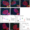
Fig 3. Tamoxifen reduces β-cell proliferation in mixed genetic background mice.
(a-e) Representative islet images for vehicle (a) and increasing doses of tamoxifen (b-e) stained for insulin (red), BrdU (green), and DAPI (blue). Scale bar: 100μm. (f) Cumulative β-cell proliferation of BrdU+ insulin+ cells after continuous labeling with BrdU 2 weeks following tamoxifen treatment. (g) Cumulative acinar cell proliferation of BrdU+ acinar cells. Results expressed as mean ± SEM for 10 mice per group. (h, i) Representative islet images for vehicle (h) and tamoxifen (i) stained for insulin (red), Ki67 (green), and DAPI (blue). Scale bar: 100μm. (j) β-cell proliferation of Ki67+ insulin+ cells. *p≤0.05, **p≤0.01, ***p≤0.001, ****p≤0.001, tamoxifen (n = 7) vs. vehicle (n = 6).
Fig 4
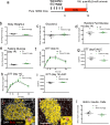
Fig 4. Tamoxifen increases fasting glucose but improves glucose tolerance in pure 129SF1/J mice; Tamoxifen does not significantly reduce β-cell proliferation in pure 129SF1/J mice.
(a) Schematic indicating time course of treatment. 129SF1/J mice received daily oral gavage with tamoxifen (0.2mg/g body weight) or vehicle for 5 days. Mice were sequentially labeled with BrdU for two weeks each, starting immediately after tamoxifen treatment. Body weight (b) and blood glucose (c) change from the start of treatment (day -5) to the end of the experiment (day 14). Random fed (d) and fasting (e) blood glucose levels at the end of the experiment. (f, g) GTT curves (f) and GTT AUC calculations (g) 5 days after tamoxifen treatment. (h, i) GTT curves (h) and GTT AUC (i) calculations for 14 days after tamoxifen treatment. (j, k) Representative islet images for vehicle (j) and tamoxifen (k) stained for insulin (red), BrdU (green), and DAPI (blue). Scale bar: 100μm. (l) Cumulative β-cell proliferation of BrdU+ insulin+ cells after continuous labeling with BrdU 2 weeks following tamoxifen treatment. Results expressed as mean ± SEM for 5 controls and 3 tamoxifen-treated mice per group. *p≤0.05, **p≤0.01, tamoxifen vs. vehicle.
Fig 5
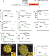
Fig 5. Tamoxifen decreases fasting glucose and improves glucose tolerance in C57Bl/6J mice; Tamoxifen significantly reduces β-cell proliferation in C57Bl/6J mice.
(a) Schematic indicating time course of treatment. C57Bl/6J mice received daily oral gavage with tamoxifen (0.2mg/g body weight) or vehicle for 5 days. Mice were sequentially labeled with BrdU for two weeks each, starting immediately after tamoxifen treatment. (b, c) Body weight (b) and blood glucose (c) change from the start of treatment (day 0) to the end of the experiment (day 14). Random fed (d) and fasting (e) blood glucose levels at the end of the experiment. (f, g) GTT curves (f) and GTT AUC calculations (g) for 5 days after tamoxifen treatment. (h, i) GTT curves (h) and GTT AUC (i) calculations for 14 days after tamoxifen treatment. (j, k) Representative islet images for vehicle (j) and tamoxifen (k) stained for insulin (red), BrdU (green), and DAPI (blue). Scale bar: 100μm. (l) Cumulative β-cell proliferation of BrdU+ insulin+ cells after continuous labeling with BrdU 2 weeks following tamoxifen treatment. Results expressed as mean ± SEM for 5 controls and 5 tamoxifen-treated mice per group. **p≤0.01, ***p≤0.001, tamoxifen vs. vehicle.
Fig 6
Fig 6. Altered gene expression in islets harvested from tamoxifen-treated mixed genetic background (F1 hybrid B6129SF1/J) mice.
(a) Schematic indicating time course of treatment. (b) RT-PCR on islet cDNA from tamoxifen-treated mice. Several common genes known to influence cell cycle entry of β-cells were chosen for analysis, including various cyclins, cyclin inhibitors, and tumor suppressors. Results expressed as mean ± SEM for 5 controls and 5 tamoxifen-treated mice per group. *p≤0.05, **p≤0.01, ***p≤0.001, tamoxifen vs. vehicle.

Similar articles

Cited by

References

    1. Kushner JA. The role of aging upon beta cell turnover. J Clin Invest. 2013;123(3):990–5. 10.1172/JCI64095 - DOI - PMC - PubMed
    1. Dor Y, Brown J, Martinez OI, Melton DA. Adult pancreatic beta-cells are formed by self-duplication rather than stem-cell differentiation. Nature. 2004;429(6987):41–6. 10.1038/nature02520 . - DOI - PubMed
    1. Teta M, Rankin MM, Long SY, Stein GM, Kushner JA. Growth and regeneration of adult beta cells does not involve specialized progenitors. Dev Cell. 2007;12(5):817–26. 10.1016/j.devcel.2007.04.011 . - DOI - PubMed
    1. Salpeter SJ, Khalaileh A, Weinberg-Corem N, Ziv O, Glaser B, Dor Y. Systemic regulation of the age-related decline of pancreatic beta-cell replication. Diabetes. 2013;62(8):2843–8. 10.2337/db13-0160 - DOI - PMC - PubMed
    1. Riley KG, Pasek RC, Maulis MF, Peek J, Thorel F, Brigstock DR, et al. CTGF modulates adult beta-cell maturity and proliferation to promote beta-cell regeneration in mice. Diabetes. 2014;64(4):1264–98. 10.2337/db14-1195 . - DOI - PMC - PubMed

Publication types