. 2019 Sep 6;14(9):e0222158.
doi: 10.1371/journal.pone.0222158. eCollection 2019.

Possible roles of monocytes/macrophages in response to elephant endotheliotropic herpesvirus (EEHV) infections in Asian elephants (Elephas maximus)

Affiliations

Possible roles of monocytes/macrophages in response to elephant endotheliotropic herpesvirus (EEHV) infections in Asian elephants (Elephas maximus)

Saralee Srivorakul et al. PLoS One. .

Abstract

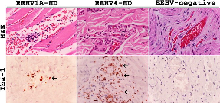
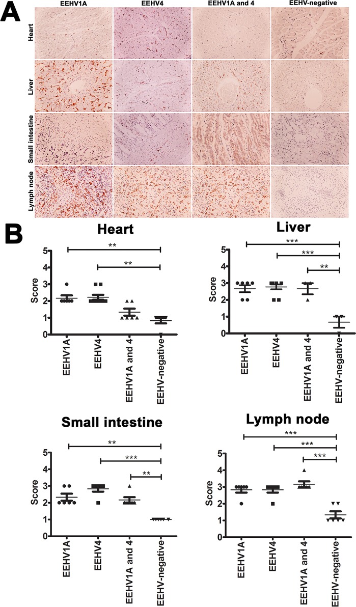
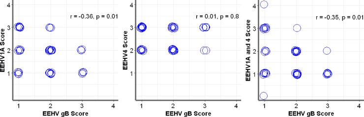
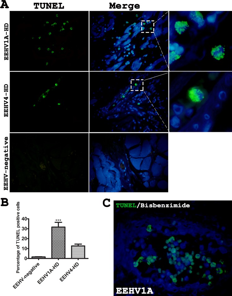
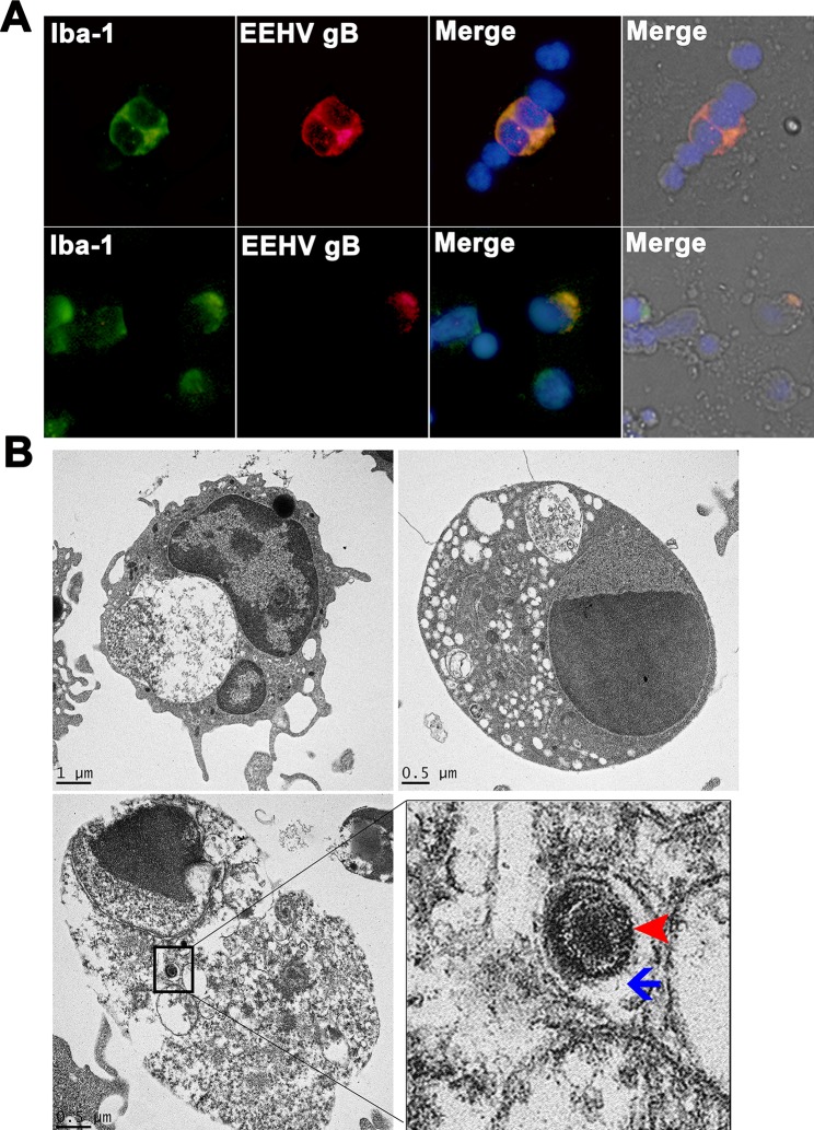
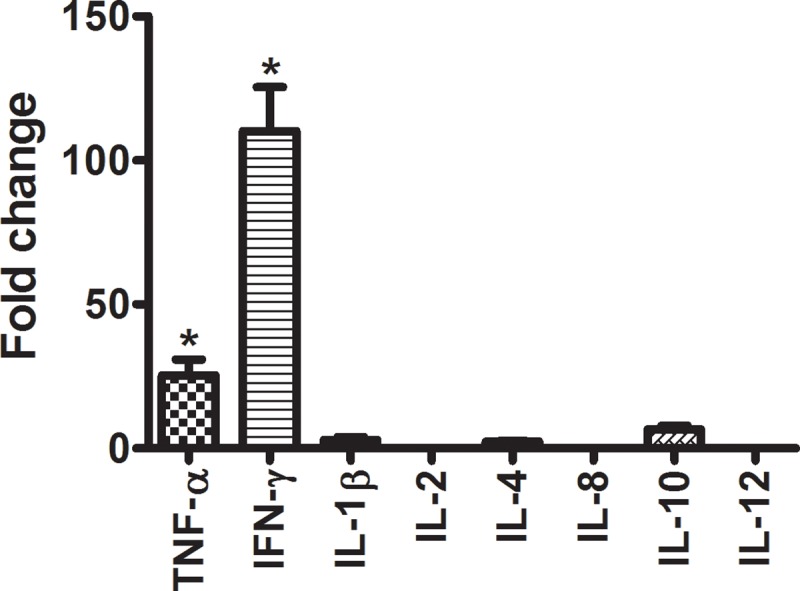
Elephant endotheliotropic herpesvirus-hemorrhagic disease (EEHV-HD) is the primary cause of acute, highly fatal, hemorrhagic diseases in young Asian elephants. Although monocytopenia is frequently observed in EEHV-HD cases, the role monocytes play in EEHV-disease pathogenesis is unknown. This study seeks to explain the responses of monocytes/macrophages in the pathogenesis of EEHV-HD. Samples of blood, frozen tissues, and formalin-fixed, paraffin-embedded (FFPE) tissues from EEHV1A-HD, EEHV4-HD, co-infected EEHV1A and 4-HD, and EEHV-negative calves were analyzed. Peripheral blood mononuclear cells (PBMCs) from the persistent EEHV4-infected and EEHV-negative calves were also studied. The results showed increased infiltration of Iba-1-positive macrophages in the inflamed tissues of the internal organs of elephant calves with EEHV-HD. In addition, cellular apoptosis also increased in the tissues of elephants with EEHV-HD, especially in the PBMCs, compared to the EEHV-negative control. In the PBMCs of persistent EEHV4-infected elephants, cytokine mRNA expression was high, particularly up-regulation of TNF-α and IFN-γ. Moreover, viral particles were observed in the cytoplasm of the persistent EEHV4-infected elephant monocytes. Our study demonstrated for the first time that apoptosis of the PBMCs increased in cases of EEHV-HD. Furthermore, this study showed that monocytes may serve as a vehicle for viral dissemination during EEHV infection in Asian elephants.


Conflict of interest statement

The authors have declared that no competing interests exist.

Figures

Fig 1
Fig 1. Histopathological and immunohistochemical labeling for Iba-1 of tissue samples from the EEHV1A-HD, EEHV4-HD and EEHV-negative calves.
EEHV infections resulted in a non-suppurative and granulomatous vasculitis and perivasculitis, and increased extravasation of the Iba-1 positive cells (arrows) out of blood vessels of the EEHV-HD calves, when compared to the EEHV-negative control.
Fig 2
Fig 2. Immunohistochemical labeling and scoring of Iba-1 positive cells in tissues of the EEHV1A-HD, EEHV4-HD, co-infected EEHV1A and 4-HD, and EEHV-negative calves.
Significant immunolabeling of Iba-1 antibodies was observed within the blood vessels and parenchymal tissues of various internal organs of EEHV-infected calves, including the hearts, livers, small intestines and lymph nodes, compared to the EEHV-negative control group (A, B). Scoring was obtained from three independent observers and data presented as a mean ± standard error. Asterisks indicate statistical significance (*p<0.05, **p<0.01, ***p<0.001), compared to the EEHV-negative control group.
Fig 3
Fig 3. Correlative scoring of EEHV gB-positive and Iba-1-positive cells using Spearman’s rank correlation test.
Immunolabeling positive cells of the EEHV gB and Iba-1 antibodies from the hearts, lungs, livers, lymph nodes, kidneys, salivary glands, and intestines were scored by three independent observers. Scoring of EEHV gB positive and Iba-1positive cells in the EEHV1A-HD and co-infected EEHV1A and 4-HD calves revealed a significant negative correlation; the Iba-1 positive cells in the tissues increased while only a low level of EEHV gB positive cells were detected. No significant correlation was observed in the EEHV4-HD calves.
Fig 4
Fig 4. Terminal deoxynucleotidyl transferase-mediated dUTP nick end labelling (TUNEL) assay of the tissues from the EEHV1A-HD, EEHV4-HD and EEHV-negative calves.
TUNEL assay revealed remarkable and significant apoptosis in the EEHV-HD calves (A, B). Apoptosis in the EEHV1A-HD calves was predominantly seen in the mononuclear cells within the blood vessels, compared to the EEHV-negative control (C). Data presented as mean ± standard error. Asterisks indicate statistical significance (***p<0.001) compared to the EEHV-negative control group.
Fig 5
Fig 5. Immunofluorescence and electron micrograph of the persistent EEHV4-infected elephant PBMCs.
PBMCs were obtained from healthy elephants known to be infected with EEHV4 for at least six months. PBMCs were then either double immunofluorescent stained with anti-EEHV gB and anti-Iba-1 antibodies (A) or processed for transmission electron microscopy (B). Staining of elephant PBMCs with anti-EEHV gB (red) revealed antigen distribution in the cytoplasm of the Iba-1 positive cells (green), suggesting their monocytic phenotype (A). The EEHV infection was observed in both non-degenerated and degenerated Iba-1 positive cells. Transmission electron micrographs of the EEHV4-positive elephant monocytes revealed segmented nuclei and variable degrees of vacuolation in the cytoplasm, with some endosomes containing virus particles (inset, B). Degenerated monocytes were observed through chromatin and cytoplasmic lysis. Enveloped viruses (arrowheads) with a diameter of ~150 nm were observed in the cytoplasmic endosomes (arrows) of degenerated cells (B).
Fig 6
Fig 6. Upregulation of cytokine mRNA expression in the persistent EEHV4-infected calves.
Quantification of cytokine mRNA expressions of elephant PBMCs revealed significant up-regulations of TNF-α and IFN-γ in the persistent EEHV4-infected calves compared to the EEHV-negative controls. Data represent the mean ± standard error from two independent experiments. Asterisks indicate statistically significant differences (*p<0.05) compared to the EEHV-negative controls.

Similar articles

Cited by

References

    1. Long SY, Latimer EM, Hayward GS. Review of elephant endotheliotropic herpesviruses and acute hemorrhagic disease. ILAR Journal. 2016;56(3):283–96. 10.1093/ilar/ilv041 - DOI - PMC - PubMed
    1. Wilkie GS, Davison AJ, Kerr K, Stidworthy MF, Redrobe S, Steinbach F, et al. First fatality associated with elephant endotheliotropic herpesvirus 5 in an Asian elephant: pathological findings and complete viral genome sequence. Scientific Reports. 2014. September 09;4:6299 10.1038/srep06299 - DOI - PMC - PubMed
    1. Richman LK, Montali RJ, Garber RL, Kennedy MA, Lehnhardt J, Hildebrandt T, et al. Novel endotheliotropic herpesviruses fatal for Asian and African elephants. Science. 1999. February 19;283(5405):1171–6. 10.1126/science.283.5405.1171 - DOI - PubMed
    1. Richman LK, Zong JC, Latimer EM, Lock J, Fleischer RC, Heaggans SY, et al. Elephant endotheliotropic herpesviruses EEHV1A, EEHV1B, and EEHV2 from cases of hemorrhagic disease are highly diverged from other mammalian herpesviruses and may form a new subfamily. Journal of Virology. 2014. December;88(23):13523–46. 10.1128/JVI.01673-14 - DOI - PMC - PubMed
    1. Dastjerdi A, Seilern-Moy K, Darpel K, Steinbach F, Molenaar F. Surviving and fatal Elephant Endotheliotropic Herpesvirus-1A infections in juvenile Asian elephants—lessons learned and recommendations on anti-herpesviral therapy. BMC Veterinary Research. 2016. August 27;12(1):178 10.1186/s12917-016-0806-5 - DOI - PMC - PubMed

Publication types