Review
. 2019 Apr:144:148-161.
doi: 10.1016/j.addr.2019.08.010. Epub 2019 Sep 3.

Domesticating the foreign body response: Recent advances and applications

Affiliations
Review

Domesticating the foreign body response: Recent advances and applications

Omid Veiseh et al. Adv Drug Deliv Rev. 2019 Apr.

Abstract

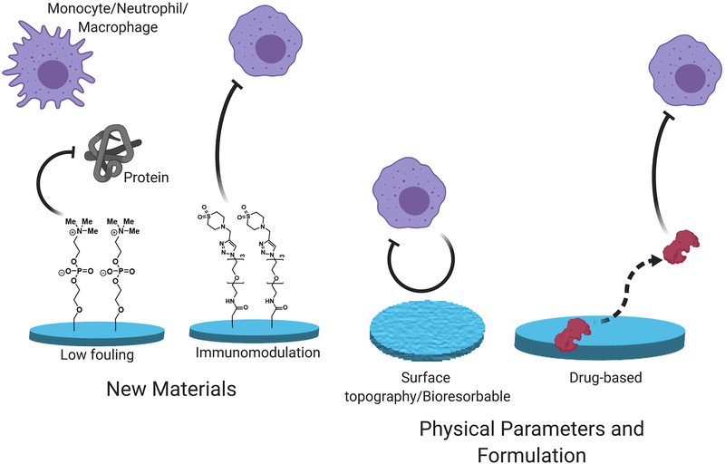
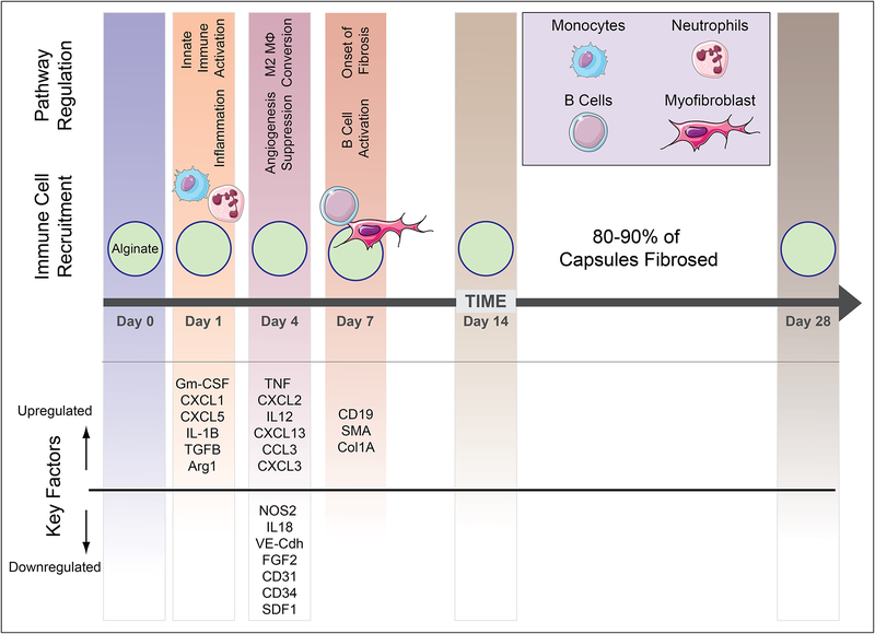
The foreign body response is an immunological process that leads to the rejection of implanted devices and presents a fundamental challenge to their performance, durability, and therapeutic utility. Recent advances in materials development and device design are now providing strategies to overcome this immune-mediated reaction. Here, we briefly review our current mechanistic understanding of the foreign body response and highlight new anti-FBR technologies from this decade that have been applied successfully in biomedical applications relevant to implants, devices, and cell-based therapies. Further development of these important technologies promises to enable new therapies, diagnostics, and revolutionize the management of patient care for many intractable diseases.

Keywords: Alginate; Biomaterials; Cell encapsulation; Devices; Diabetes; Fibrosis; Foreign body response; Hydrogel; Immunology; Immunomodulation; Implantation; Zwitterionic.


Conflict of interest statement

Disclosure

The authors of this manuscript have conflicts of interest to disclose as described by Advanced Drug Delivery Reviews. O.V. and A.J.V. are scientific founders of Sigilon Therapeutics.

Figures

Figure 1.
Figure 1.
An overview of the approaches taken to mitigate the immunological processes responsible for the foreign body response (FBR).
Figure 2.
Figure 2.
Kinetic profile of host immune response to implanted biomaterials based on interpretation of data presented references [24,25].
Figure 3.
Figure 3.
Systems profiling of immune cell perturbations and its influence on Foreign Body Reactions and fibrosis. Using a combination of genetic knockouts and targeted immune depletions, macrophages were the only immune cell population required for fibrotic encapsulation of implanted alginate spheres. Right column: representative summary responses based on phase contrast images showing fibrosis levels on 500 μm alginate spheres retrieved from wild type C57BL/6 mice (n = 5/group), after 14-day intraperitoneal implantations. * = as reported. N/A = while available, not tested due to not being essential players. Figure and caption adapted with permission from reference [24].
Figure 4.
Figure 4.
Extravasation of macrophage cells from peripheral tissue on to spherical materials of 0.5 mm and 1.5 mm spheres. (a) schematic describing how smaller spheres better conform to peripheral adjacent tissue cervices compared to larger spheres and this influence on the ability of immune cells to extravasate on to implants from vasculature. In vivo intravital imaging of macrophage behavior and accumulation at 7 days post-implantation on to (b) 0.5 mm and (c) 1.5 mm diameter sized Ba+ crosslinked alginate spheres. (macrophages depicted in green, peripheral tissue in white and implanted spheres in magenta). Figure and caption adapted with permission from reference [25].
Figure 5.
Figure 5.
Silicone breast implant embedded with Pirfenidone. (a) Both smooth and textured implants were tested in rats. (b) Fibrosis analysis of the implants based on Masson’s trichrome staining of immunohistological sections. Drug-treated mice showed reduced fibrosis to control implants. Images of (c) smooth, (d) textured, (e) drug-treated smooth, and (f) drug-treated textured implants. Figure and caption adapted with permission from reference [93].
Figure 6.
Figure 6.
A bioresorbable fibre optic probe for making physiological measurements. (a) The Si nanomembrane photodetector is encapsulated in a PLGA-based fibre optic fibre. (b) Computed tomography imaging in mice showing the gradual resorption of the probe over a period of 7 weeks. (c) In vivo function of the bioresorbable spectrometer implanted in mice, showing measurement of brain temperature during food intake tests. Figure and caption adapted with permission from reference [106].
Figure 7.
Figure 7.
Low-fouling polymeric coatings improve CGM performance. (a) Zwitterionic Poly(MPC) polymers were copolymerized with thiol-containing monomers. (b) A dopamine-based strategy was used to coat CGM electrodes. Glucose-sensing performance of the coated electrodes was superior in both (c) murine and (d) NHP models of diabetes. Figure and caption adapted with permission from reference [115].
Figure 8.
Figure 8.
Small-molecule modifications of the alginate backbone can mitigate the FBR. (a) The lead small-molecule modifications that were identified from a combinatorial modified alginate library. These lead modified alginates successfully mitigated fibrosis in the IP space of both immunocompetent murine and NHP animal models. (b) Representative phase contrast imaging of retrieved capsules for the control (SLG20) and modified alginate formulations after 4 weeks in NHP are shown. (c) Immunofluorescence imaging of the retrieved capsules in (b) showed reduced markers of general cellular material (DAPI), myofibroblasts (α-SMA), and macrophages (CD11b). Figure and caption adapted with permission from reference [117].
Figure 9.
Figure 9.
Modified alginates enable successful long-term transplantation of human-derived SC-β cells in diabetic, immunocompetent mice. (a) Encapsulated SC- β cells in TMTD-alginate spheres with 1.5mm diameters. (b) The modified alginate spheres supported long-term (6 month) glycemic correction. The encapsulated cells showed signs of reduced fibrosis even at the end of the 6-month period. (c) Brightfield imaging of a retrieved sphere with encapsulated SC-β cell cluster seen inside. (d) Immunofluorescence straining, (e) Masson’s trichrome, and (f) H&E histology of the retrieved spheres. Figure and caption adapted with permission from reference [119].
Figure 10.
Figure 10.
Schematic of a macroscaled device design that combines various approaches to addressing foreign body response to achieve improved in vivo performance for cell-based therapies.

References

    1. Anderson JM, Rodriguez A, Chang DT, Foreign body reaction to biomaterials, Seminars in Immunology. 20 (2008) 86–100. doi:10.1016/j.smim.2007.11.004. - DOI - PMC - PubMed
    1. Wick G, Grundtman C, Mayerl C, Wimpissinger T-F, Feichtinger J, Zelger B, Sgonc R, Wolfram D, The Immunology of Fibrosis, Annual Review of Immunology. 31 (2013) 107–135. doi:10.1146/annurev-immunol-032712-095937. - DOI - PubMed
    1. Wynn TA, Ramalingam TR, Mechanisms of fibrosis: therapeutic translation for fibrotic disease, Nat Med. 18 (2012) 1028–1040. doi:10.1038/nm.2807. - DOI - PMC - PubMed
    1. Ward WK, A Review of the Foreign-body Response to Subcutaneously-implanted Devices: The Role of Macrophages and Cytokines in Biofouling and Fibrosis, J Diabetes Sci Technol. 2 (2008) 768–777. - PMC - PubMed
    1. Zhang L, Cao Z, Bai T, Carr L, Ella-Menye J-R, Irvin C, Ratner BD, Jiang S, Zwitterionic hydrogels implanted in mice resist the foreign-body reaction, Nat Biotech. 31 (2013) 553–556. doi:10.1038/nbt.2580. - DOI - PubMed

Publication types