. 2019 Sep 5;20(18):4345.
doi: 10.3390/ijms20184345.

Functional Dissection of pri-miR-290~295 in Dgcr8 Knockout Mouse Embryonic Stem Cells

Affiliations

Functional Dissection of pri-miR-290~295 in Dgcr8 Knockout Mouse Embryonic Stem Cells

Ming Shi et al. Int J Mol Sci. .

Abstract

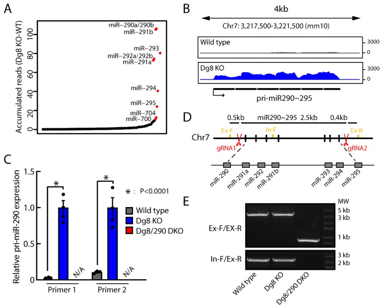
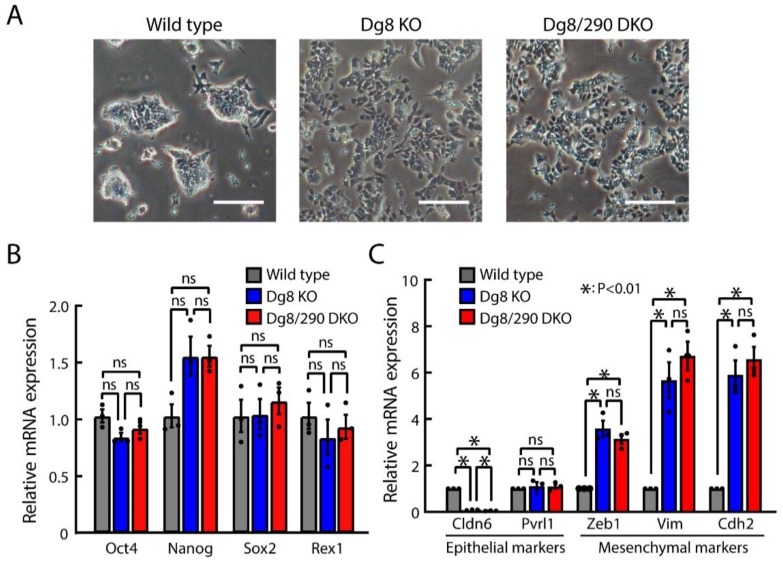
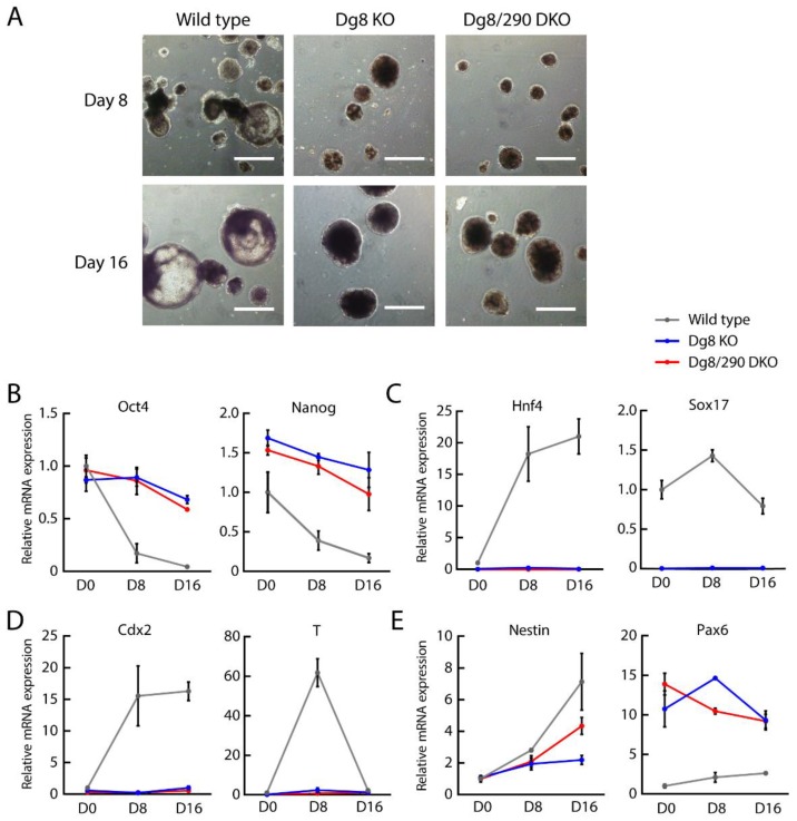
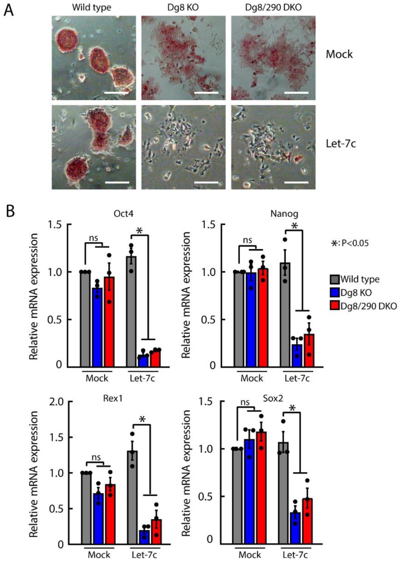
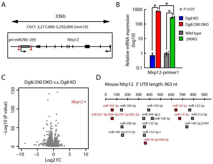
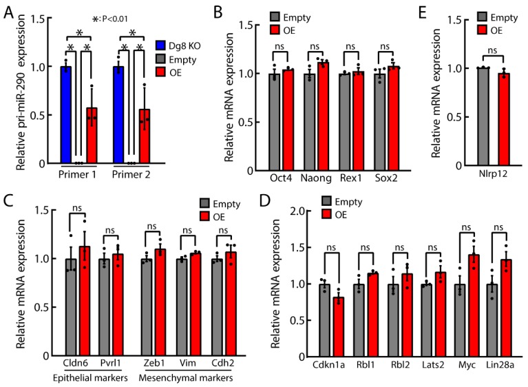
The DiGeorge syndrome critical region gene 8 (Dgcr8) knockout strategy has been widely used to study the function of canonical microRNAs (miRNAs) in vitro and in vivo. However, primary miRNA (pri-miRNA) transcripts are accumulated in Dgcr8 knockout cells due to interrupted processing. Whether abnormally accumulated pri-miRNAs have any function is unknown. Here, using clustered regularly interspaced short palindromic repeats system/CRISPR-associated protein 9 (CRISPR/Cas9), we successfully knocked out the primary microRNA-290~295 (pri-miR-290~295) cluster, the most highly expressed miRNA cluster in mouse embryonic stem cells (ESCs), in Dgcr8 knockout background. We found that the major defects associated with Dgcr8 knockout in mouse ESCs, including higher expression of epithelial-to-mesenchymal transition (EMT) markers, slower proliferation, G1 accumulation, and defects in silencing self-renewal, were not affected by the deletion of pri-miR-290~290 cluster. Interestingly, the transcription of neighboring gene nucleotide-binding oligomerization domain, leucine rich repeat and pyrin domain containing 12(Nlrp12) was upregulated upon the deletion of the pri-miR-290~295 cluster. Together, our results suggested that the major defects in Dgcr8 knockout ESCs were not due to the accumulation of pri-miR-290~295, and the deletion of miRNA genes could affect the transcription of neighboring DNA elements.

Keywords: DiGeorge syndrome critical region gene 8; Nlrp12; cell cycle; differentiation; embryonic stem cells; miR-290~295 cluster; pluripotency; primary microRNA; self-renewal.


Conflict of interest statement

The authors declare no conflict of interest.

Figures

Figure 1
Figure 1
CRISPR/Cas9 mediated pri-miR-290~295 knock out in Dgcr8 knockout embryonic stem cells (ESCs). (A) Accumulated reads mapped to pre-miRNA loci. Normalized reads per million total reads are shown. (B) RNA-Seq tracks for pri-miR-290~295 locus in wild type and Dgcr8 knockout ESCs. Shown are normalized read counts per million. (C) Pri-miR-290~295 levels in ESCs measured by RT-qPCR. Results from two different primer pairs targeting pri-miR-290 were exhibited. Shown are means ± SEM, n = 3 independent experiments. The p-value was determined by one-way ANOVA followed by two-tailed Tukey’s test. (D) A diagram illustrating the genomic locus of the miR-290~295 cluster and the knockout strategy. Locations of the targeting sequences for the gRNAs and of the primers for PCR verification are indicated by red scissors and yellow squares. (E) Genomic PCR confirming the deletion of miR-290 fragment using different primers, as shown in Figure 1C. Dg8 KO, Dgcr8 knockout; Dg8/290 DKO, Dgcr8/pri-miR-290~295 double knockout.
Figure 2
Figure 2
Pri-miR-290~295 transcripts have no role in regulating pluripotency and epithelial-to-mesenchymal transition (EMT)-like property in ESCs. (A) Morphology of ESC colonies under standard culture conditions. Representative images from different groups are shown. Scale bars = 200 μm. (B,C) mRNA levels of pluripotency marker genes (B) and EMT-associated genes (C) were analyzed by RT-qPCR. The β-actin gene was taken as an internal control. For each gene, data were normalized to the mRNA level of wild type ESCs. Shown are mean ± SEM, n = 3 independent experiments. The p-value was determined by one-way ANOVA followed by two-tailed Tuke’s test. Dg8 KO, Dgcr8 knockout; Dg8/290 DKO, Dgcr8/pri-miR-290~295 double knockout.
Figure 3
Figure 3
The accumulation of pri-miR-290~295 transcripts has no role in proliferation and cell cycle defects of Dgcr8 knockout ESCs. (A) Cell population doubling time was calculated during serial passages in ESC culture. (B,C) Cell cycle profile with propidium iodide (PI) staining of ESCs was analyzed by flow cytometry, and the fraction of cells in the various phases of the cell cycle was calculated. (D) Expression of cell cycle genes in ESCs was determined by RT-qPCR. The β-actin gene was taken as an internal control. For each gene, data were normalized to the mRNA level of wild type ESCs. Shown are mean ± SEM, n = 3 independent experiments. The p-value was determined by one-way ANOVA followed by two-tailed Tukey’s test. Dg8 KO, Dgcr8 knockout; Dg8/290 DKO, Dgcr8/pri-miR-290~295 double knockout.
Figure 4
Figure 4
The accumulation of pri-miR-290~295 transcripts has no role for embryoid body (EB) differentiation defects of Dgcr8 knockout ESCs. (A) Representative images of embryoid bodies from ESCs with a different background. Scale bars = 500 μm. (BE) Expression of pluripotency and differentiation markers at day 0, 8, and 16 of EB differentiation. Expression of representative markers of pluripotency (Oct4, Nanog), endoderm (hepatic nuclear factor 4 alpha (Hnf4), Ssex determining region Y-Box 17 (Sox17)), mesoderm (T-box transcription factor T brachyury (T), caudal type homeobox 2 (Cdx2)), and ectoderm (Pax6, Nestin) was measured by RT-qPCR. The β-actin gene was used as an internal control. For each gene, data were normalized to the mRNA level of wild type ESCs (D0). Shown are mean ± SEM, n = 3 independent experiments. The p-value was determined by one-way ANOVA followed by two-tailed Tukey’s test. Dg8 KO, Dgcr8 knockout; Dg8/290 DKO, Dgcr8/pri-miR-290~295 double knockout.
Figure 5
Figure 5
The accumulation of pri-miR-290~295 has no role in let-7 mediated silencing of self-renewal of Dgcr8 knockout ESCs. (A) Representative images of ESC colonies with alkaline phosphatase staining after mock or let-7c transfection. Scale bars = 200 μm. (B) qRT-PCR analysis of self-renewal genes in ESCs after mock or let-7c transfection. The β-actin gene was used as a control. For each gene, data were normalized to the mRNA level of wild type ESCs. Shown are means ± SEM, n = 3 independent experiments. The p-value was determined by one-way ANOVA followed by two-tailed Tukey’s test. Dg8 KO, Dgcr8 knockout; Dg8/290 DKO, Dgcr8/pri-miR-290~295 double knockout.
Figure 6
Figure 6
Deletion of pri-miR-290~295 cluster leads to remarkable upregulation of Nlrp12 in both wild type and Dgcr8 knockout ESCs. (A) A diagram illustrating the genomic locus of miR-290~295 cluster and Nlrp12. Caspase 9 (Cas9) cleavage positions are indicated by red scissors. (B) qRT-PCR analysis for mRNA expression of Nlrp12. The β-actin gene was used as a control. For each gene, data were normalized to the mRNA level of wild type ESCs. Shown are mean ± SEM, n = 3 biological replicates. The p-value was determined by one-way ANOVA followed by two-tailed Tukey’s test. Dg8 KO, Dgcr8 knockout; Dg8/290 DKO, Dgcr8/pri-miR-290~295 double knockout; 290KO, pri-miR-290~295 knockout. (C) Volcano plot representation of differential expression analysis of genes in Dgcr8/pri-miR-290~295 double knockout versus Dgcr8 knockout ESCs. The x-axis shows log2 fold changes, and the y-axis shows log10 p values. (D) Predicted miRNA binding sites in Nlrp12 3′ untranslated region by Targetscan. miRNAs expressed more than 100 copy per cell in wild type ESCs are labeled in red.
Figure 7
Figure 7
Transgene expression of pri-miR-290~295 has no impact on the expression of pluripotency, EMT- and cell cycle-related genes, and Nlrp12. (A) RT-qPCR analysis for pri-miR-290~295 in Dgcr8 knockout, Dgcr8/pri-miR-290~295 knockout ESCs transfected with empty control or vectors overexpressing pri-miR-290~295. The overexpression in double knockout ESCs restored the level of pri-miR-290~295 to ~60% of that in Dgcr8 knockout ESCs. (B) RT-qPCR analysis for mRNA expression of pluripotency genes. (C) RT-qPCR analysis for mRNA expression of EMT related genes. (D) RT-qPCR analysis for mRNA expression of cell cycle-related genes. (E) RT-qPCR analysis for mRNA expression of Nlrp12. For all panels, the β-actin gene was used as a control. For each gene, data were normalized to the mRNA level of Dgcr8/pri-miR-290~295 knockout ESCs transfected with empty control except in A, in which data were normalized to Dgcr8 knockout ESCs. Shown are mean ± SEM, n = 3 biological replicates. The p-value was determined by one-way ANOVA followed by two-tailed Tukey’s test in A and by two-tailed unpaired Student’s t-test in Figure 7B–E. Dg8 KO, Dgcr8 knockout; Empty, Dgcr8/pri-miR-290~295 double knockout ESCs transfected with empty control overexpression vectors; OE, Dgcr8/pri-miR-290~295 double knockout ESCs transfected with pri-miR-290~295 overexpression vectors.

Similar articles

Cited by

References

    1. Bartel D.P. Metazoan MicroRNAs. Cell. 2018;173:20–51. doi: 10.1016/j.cell.2018.03.006. - DOI - PMC - PubMed
    1. Fischer S., Handrick R., Aschrafi A., Otte K. Unveiling the principle of microRNA-mediated redundancy in cellular pathway regulation. RNA Biol. 2015;12:238–247. doi: 10.1080/15476286.2015.1017238. - DOI - PMC - PubMed
    1. Guo W.T., Wang Y. Dgcr8 knockout approaches to understand microRNA functions in vitro and in vivo. Cell. Mol. Life Sci. 2019;76:1697–1711. doi: 10.1007/s00018-019-03020-9. - DOI - PMC - PubMed
    1. Murchison E.P., Partridge J.F., Tam O.H., Cheloufi S., Hannon G.J. Characterization of Dicer-deficient murine embryonic stem cells. Proc. Natl. Acad. Sci. USA. 2005;102:12135–12140. doi: 10.1073/pnas.0505479102. - DOI - PMC - PubMed
    1. Kanellopoulou C., Muljo S.A., Kung A.L., Ganesan S., Drapkin R., Jenuwein T., Livingston D.M., Rajewsky K. Dicer-deficient mouse embryonic stem cells are defective in differentiation and centromeric silencing. Genes Dev. 2005;19:489–501. doi: 10.1101/gad.1248505. - DOI - PMC - PubMed

LinkOut - more resources