. 2019 Sep 5;11(9):824.
doi: 10.3390/v11090824.

Erythroid Progenitor Cells in Atlantic Salmon (Salmo salar) May Be Persistently and Productively Infected with Piscine Orthoreovirus (PRV)

Affiliations

Erythroid Progenitor Cells in Atlantic Salmon (Salmo salar) May Be Persistently and Productively Infected with Piscine Orthoreovirus (PRV)

Muhammad Salman Malik et al. Viruses. .

Abstract

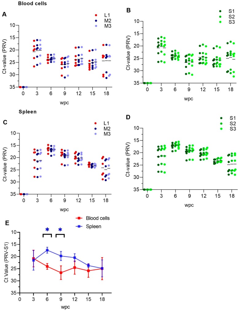
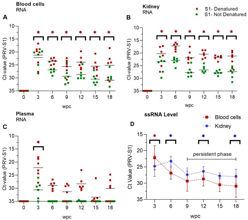
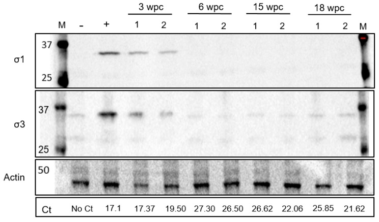
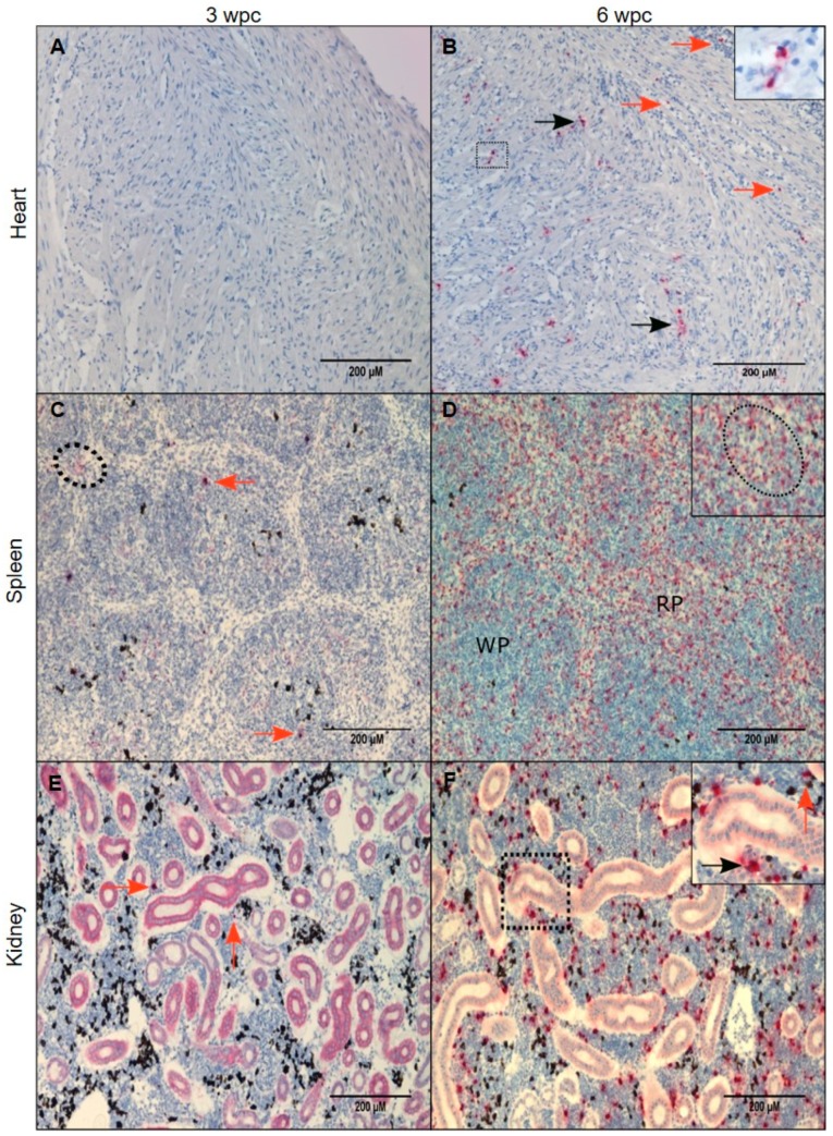
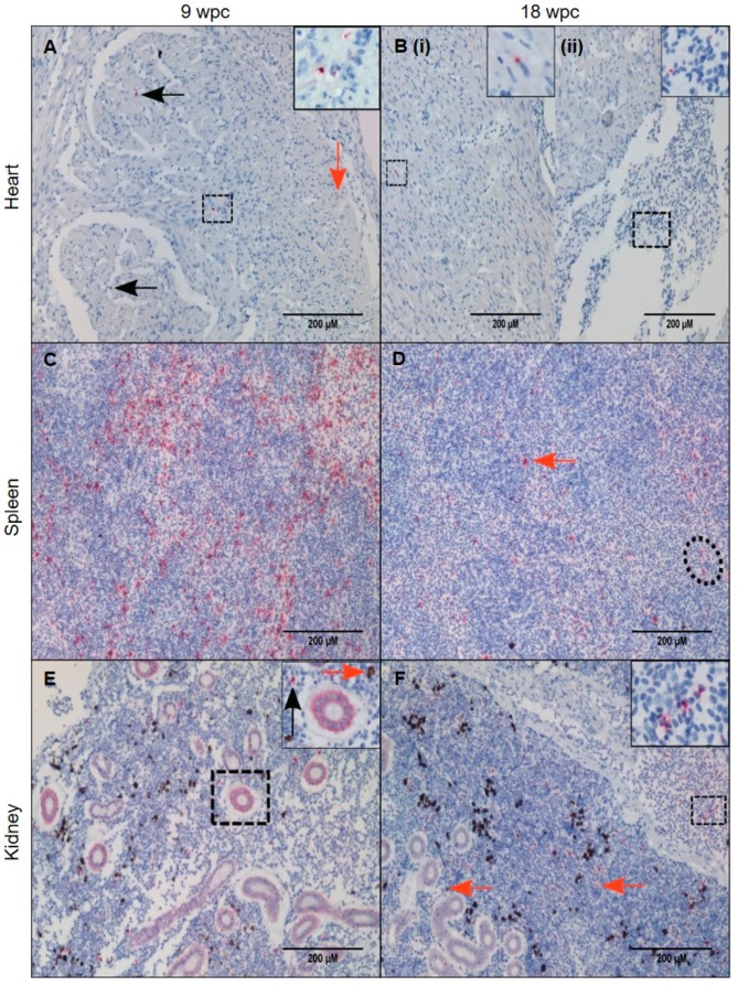
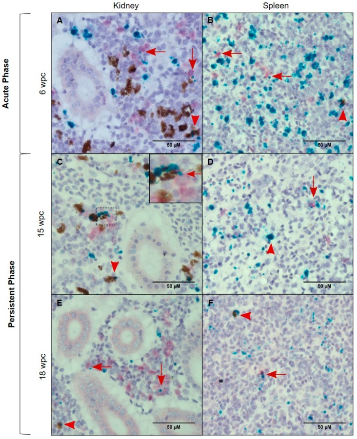
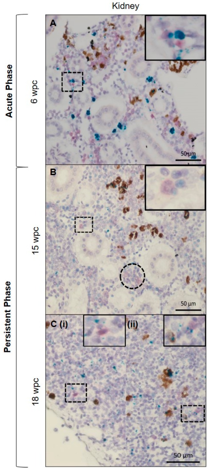
Piscine orthoreovirus (PRV-1) can cause heart and skeletal muscle inflammation (HSMI) in farmed Atlantic salmon (Salmo salar). The virus targets erythrocytes in the acute peak phase, followed by cardiomyocytes, before the infection subsides into persistence. The persistent phase is characterized by high level of viral RNA, but low level of viral protein. The origin and nature of persistent PRV-1 are not clear. Here, we analyzed for viral persistence and activity in various tissues and cell types in experimentally infected Atlantic salmon. Plasma contained PRV-1 genomic dsRNA throughout an 18-week long infection trial, indicating that viral particles are continuously produced and released. The highest level of PRV-1 RNA in the persistent phase was found in kidney. The level of PRV-1 ssRNA transcripts in kidney was significantly higher than that of blood cells in the persistent phase. In-situ hybridization assays confirmed that PRV-1 RNA was present in erythroid progenitor cells, erythrocytes, macrophages, melano-macrophages and in some additional un-characterized cells in kidney. These results show that PRV-1 establishes a productive, persistent infection in Atlantic salmon and that erythrocyte progenitor cells are PRV target cells.

Keywords: PRV-1; erythroid progenitor cells; persistence; piscine orthoreovirus.


Conflict of interest statement

The authors declare no conflict of interest.

Figures

Figure 1
Figure 1
Expression analysis of PRV-1 segments and transcripts. qPCR detection of segment L1, M2, M3, S1-S3 and corresponding transcripts in blood cells (A,B) and spleen (C,D) from Atlantic salmon. Individual fish shown as dots, and mean Ct value-PRV as lines. n = 6 for each point. (E) S1 as a representative for PRV genomic segments expression between blood cells and spleen. Paired analysis was performed by non-parametric Wilcoxon matched pairs signed rank test (p < 0.05), asterisk (*) indicates significantly different group. wpc = weeks post challenge.
Figure 2
Figure 2
PRV-1 dsRNA and ssRNA levels. Mean Ct-values of RT-qPCR (PRV-S1) for (A) Blood cells, (B) Kidney and (C) Plasma. RNA was pre-heated (red dots) or not (green dots), indicating presence of viral dsRNA plus ssRNA transcripts or ssRNA PRV transcripts only, respectively. n = 6 for each time point. Statistical analysis between denaturated and not-denaturated samples from the same RNA was performed by non-parametric, Wilcoxon matched pairs signed rank test. Asterisk shows significantly high level (* p < 0.05) (D) Paired analysis of ssRNA Ct-values (Table S1) of kidney compared to blood cells in persistent phase (9 wpc -18 wpc). Each dot shows mean Ct-value at each time point. Statistical analysis was performed by non-parametric, Wilcoxon matched pairs signed rank test (p < 0.05), asterisk (red/blue) indicates significantly different group.). wpc = weeks post challenge.
Figure 3
Figure 3
Low levels of PRV proteins in the persistent phase. Detection of outer PRV-1 capsid proteins σ1 and σ3 in blood cells from 3–18 weeks post challenge (wpc) by western blotting. Ct values in lower row. Actin (42 kDa) was used as a protein load control. Blood cells from uninfected fish used as negative control. Positive control sample from a fish having Ct value of 17.1 and significant σ1 protein level [15].
Figure 4
Figure 4
PRV-1 localization in the acute phase. PRV-1 localization shown by in situ hybridization (red) at 3 and 6 wpc. Heart (A,B): A, (3 wpc), no PRV detection. B, (6 wpc), PRV positive cardiomyocytes in compact layer (orange arrows) and a few positive erythrocytes or infiltrating macrophage-like cells were present in the epicardium and scattered PRV staining in the spongy layer of the heart ventricle (black arrows). Spleen (C,D): C, (3 wpc), few PRV-1 positive RBCs (dotted circle) and macrophage-like cells (orange arrows). D (6 wpc) shows a large number of positively stained cells in the red pulp (RP) area. Dotted circle in magnified version in upper right corner shows cluster of cells with erythrocyte- and macrophage-like morphology. Kidney (E,F): E, (3 wpc) shows a few PRV-1 positive cells (orange arrow), primarily macrophage-like cells. F, (6 wpc), a high number of PRV-1 positive macrophage-like cells (black arrow) along with melano-macrophages (orange arrow) were found in peritubular regions of the kidney. Scale bar = 200 µm.
Figure 5
Figure 5
PRV-1 localization during the persistent phase. PRV-1 localization shown by in situ hybridization (red) at 9 and 18 wpc. Heart (A,B): A, (9 wpc) A small number of cells in the compact layer stained positive for PRV-1 (orange arrow), and similarly faintly in a few macrophage-like cells in the in the luminal space of the spongy layer (black arrows) and some circulatory cells, near blood vessel (dotted square and magnified image). B, (18 wpc) Very few cells were positive for PRV-1 with the exception of (i) some positive cardiomyocytes in the spongy layer (dotted square) or (ii) peripheral blood in the heart (dotted square). Spleen (C,D): C, (9 wpc) numerous PRV-1 positive cells in red pulp. D, (18 wpc) A few PRV-1 positive macrophage-like cells (orange arrow) and erythrocyte like cells (dotted circle). Kidney (E,F): E, (9 wpc) PRV-1 present in macrophage-like cells (black arrow) and melano-macrophages (orange arrow). F, (18 wpc) PRV-1 positive erythrocytes and macrophage-like cells throughout the section (orange arrows) and in blood vessels (magnified picture). Scale bar = 200 µm.
Figure 6
Figure 6
PRV-1 (green) localization in macrophages and melano-macrophages. Duplex in situ hybridization against PRV-1 and macrophage colony stimulating factor (MCSFR) (red). Kidney (A,C,E) and Spleen (B,D,F), showing PRV positive macrophages (red arrows) and melano-macrophages (red arrowhead) at 6 wpc, 15 wpc and 18 wpc. The use of red dye in salmon kidney sections in duplex in situ gave diffuse staining of positive cells (no background staining). The diffuse staining was different from the punctate staining seen in single in situ, or the use of green dye in duplex in situ. PRV-1 is also positive in other cell types in the sections. Scale bar = 50 µm.
Figure 7
Figure 7
PRV-1 localization in hematopoietic cells. Duplex in situ hybridization against PRV-1 (green) and erythropoietin receptor (EPOR) (red). Kidney (A) Acute phase, dually stained PRV/EPOR positive cells in hematopoietic tissue in the magnified picture. (B,C) Persistent phase, dually stained EPOR-PRV1 positive cells in hematopoietic tissue scattered around in the kidney (magnified pictures). Cluster of partially stained positive erythroid cells at 15 wpc (dotted circle). The use of red dye in salmon kidney sections in duplex in situ gave diffuse staining of positive cells (no background staining). The diffuse staining was different from the punctate staining seen in single in situ, or the use of green dye in duplex in situ. Scale bar = 50 µm.

Similar articles

Cited by

References

    1. Taranger G.L., Karlsen O., Bannister R.J., Glover K.A., Husa V., Karlsbakk E., Kvamme B.O., Boxaspen K.K., Bjorn P.A., Finstad B., et al. Risk assessment of the environmental impact of norwegian atlantic salmon farming. ICES J. Mar. Sci. 2015;72:997–1021. doi: 10.1093/icesjms/fsu132. - DOI
    1. Lovoll M., Wiik-Nielsen J., Grove S., Wiik-Nielsen C.R., Kristoffersen A.B., Faller R., Poppe T., Jung J., Pedamallu C.S., Nederbragt A.J., et al. A novel totivirus and piscine reovirus (prv) in atlantic salmon (salmo salar) with cardiomyopathy syndrome (cms) Virol. J. 2010;7:309. doi: 10.1186/1743-422X-7-309. - DOI - PMC - PubMed
    1. Palacios G., Lovoll M., Tengs T., Hornig M., Hutchison S., Hui J., Kongtorp R.T., Savji N., Bussetti A.V., Solovyov A., et al. Heart and skeletal muscle inflammation of farmed salmon is associated with infection with a novel reovirus. PLoS ONE. 2010;5:e11487. doi: 10.1371/journal.pone.0011487. - DOI - PMC - PubMed
    1. Wessel O., Braaen S., Alarcon M., Haatveit H., Roos N., Markussen T., Tengs T., Dahle M.K., Rimstad E. Infection with purified piscine orthoreovirus demonstrates a causal relationship with heart and skeletal muscle inflammation in atlantic salmon. PLoS ONE. 2017;12:e0183781. doi: 10.1371/journal.pone.0183781. - DOI - PMC - PubMed
    1. Kongtorp R.T., Taksdal T., Lyngoy A. Pathology of heart and skeletal muscle inflammation (hsmi) in farmed atlantic salmon salmo salar. Dis. Aquat. Org. 2004;59:217–224. doi: 10.3354/dao059217. - DOI - PubMed

Publication types

MeSH terms

Supplementary concepts

LinkOut - more resources