. 2019 Sep 5;8(9):1392.
doi: 10.3390/jcm8091392.

The Eye as a Window to Systemic Infectious Diseases: Old Enemies, New Imaging

Affiliations

The Eye as a Window to Systemic Infectious Diseases: Old Enemies, New Imaging

Vittorio Pirani et al. J Clin Med. .

Abstract

Background: Syphilis, tuberculosis and toxoplasmosis are major infectious diseases worldwide; all of them are multisystem pathologies and share a possible ocular involvement. In this context, a fundamental help for the definitive diagnosis is provided by the ophthalmologist, through clinical evaluation and with the aid of a multimodal imaging examination.

Methods: We hereby describe selected cases who came to our attention and were visited in our eye clinic. In all clinics, the use of retinal and optic disc multimodal imaging during ophthalmological evaluation allowed to make a diagnosis of an infectious disease.

Results: In our tertiary referral center more than 60 patients with syphilis, tuberculosis and toxoplasmosis have been evaluated in the last two years: In 60% of cases the ophthalmological evaluation was secondary to a previous diagnosis of an infectious disease, while in the remaining cases the ophthalmologist, with the help of a multimodal imaging examination and clinical evaluation, represented the physician who leads to the diagnosis.

Conclusion: Our results confirm how in these life-threatening pathologies a prompt diagnosis is mandatory and may benefit from a multidisciplinary and multimodal imaging approach, especially during ophthalmological evaluation.

Keywords: fluorescein angiography; fundus autofluorescence; indocyanine green angiography; infectious uveitis; optical coherence tomography; optical coherence tomography angiography; syphilis; toxoplasmosis; tuberculosis.


Conflict of interest statement

The authors declare no conflict of interest.

Figures

Figure 1
Figure 1
Presentation of syphilis. Fundus examination (A) shows the presence of vitreous inflammation and multiple superficial pre-retinal aggregates are evidenced on optical coherence tomography (OCT) scans (B). The patient presented a form of tertiary syphilis affecting both the eye (A,B) and the skin (C).
Figure 2
Figure 2
Follow-up OCT scans (A) showing multiple superficial pre-retinal aggregates (asterisk) and ellipsoid zone disruption (arrow) that solve after antibiotic therapy (B). Pre-retinal aggregates probably correspond to a combination of spirochetes and inflammatory cells. Ellipsoid zone disruption is probably caused by an accumulation of lipofuscin, inflammatory cells or incomplete phagocytosis of outer retinal segments.
Figure 3
Figure 3
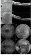
Case 1, acute syphilitic posterior placoid chorioretinitis (ASPCC). Patients affected by ASPCC show typical outer retinal abnormalities on OCT with ellipsoid zone disruption (A) (asterisk), partially resolved after the treatment (B). FA reveals early hypofluorescence and late hyperfluorescence corresponding to the lesions (C,D). ICGA shows hypocyanescence until the late stages of the examination (E,F). Green arrow: plane and orientation of the optical coherence tomography (OCT) line scan.
Figure 4
Figure 4
Case 1, Neurosyphilis. Magnetic resonance imaging (MRI) of the brain with gadolinium showing the presence of multiple hyperintense lesions in long repetition time (TR) sequences localized bilaterally in frontal hemispheres (A); in temporal, parietal and occipital left hemispheres (B) and in the right cerebellum peduncle (C). MRN: magnetic resonance neurography; R: right; L: left; P: posterior.
Figure 5
Figure 5
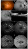
Case 2, ocular tubercles. Fundus photo (A) evidences the presence of multiple, yellowish lesions with indistinct borders. Edema around the optic disc (A) is better evidenced with B-FAF (hyperfluorescent) and OCT scan (C), showing the area of retinal pigmented epithelium (RPE) detachment (arrowheads). Contact sign over the tubercle can be seen (asterisk) on OCT (C). Granulomas are characterized by early hypo-fluorescence (D) and late hyper-fluorescence (E) after fluorescein injection. ICGA examination shows the presence of regular-shaped, hypocyanescent lesions from the early (F) to the late stages (G) of the examination.
Figure 6
Figure 6
OCTA. Areas of flow void (asterisk) in superficial (a) and deep (b) vascular plexus, corresponding to granulomas with vessels starting to grow irregularly at the edge of the lesions during the healing process.
Figure 7
Figure 7
Serpiginous-like choroiditis, clinical presentation. Lesions are usually localized around the optic disc (A) and are characterized at FA by late hypo-fluorescence (B); an area of retinal atrophy is localized next to the optic disc (A,B).
Figure 8
Figure 8
Case 3, Eales disease. Signs of vasculitis (arrowheads) are visible in the late phases of FA (A) with resolution after the therapy (B). Extended ischemic areas in the peripheral retina appear darker and without vascularization (CF).
Figure 9
Figure 9
Toxoplasma gondii chorioretinitis. Severe vitreitis with “headlight in the fog” sign; a nidus of fluffy white, necrotizing retinitis or retinochoroiditis adjacent to a variably pigmented chorioretinal scar is visible at fundus examination (A). On FA, the lesion is hypofluorescent with the presence of a hyperfluorescent border (B).
Figure 10
Figure 10
Toxoplasma gondii-related epiretinal membrane. On OCT, a hyperreflective membrane (arrowheads) can be seen adjacent to a toxoplasma scar (asterisk).
Figure 11
Figure 11
Case 4, Punctate outer retinal toxoplasmosis. OCT in the acute phase (A) showing alterations of the neurosensory retina. Hyperreflective deposits can be present within the retinal interface (A); during the follow-up, these deposits become smaller, enter the inner retinal layers and fade with time until complete resolution (B). In the active phase, the choroid is thick and hyporeflective (A) and returns to normal thickness during follow-up, becoming gradually hyperreflective (B). The presence of an epiretinal membrane (asterisk) is visible over the scarred lesion (B).
Figure 12
Figure 12
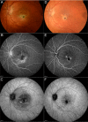
Case 4, Punctate outer retinal toxoplasmosis. Fundus examination (A) showing the presence of a yellowish lesion with a central grey spot on the macula, with indefinite borders, associated with perilesional edema. FA in the late phase displayed the presence of an enlarged foveal avascular zone (FAZ) corresponding to the area of detachment with perilesional leakage and papillitis (B). Intermediate phase ICGA evidenced a macular hypocianescent area with indefinite borders and hypercianescent striae inside the lesion (C). After the treatment, macular edema resolved (D). FA evidenced the presence of a hypofluorescent macular scar associated with RPE alterations (hyperfluorescent) in the early and late stages of the exam (E); a hypocianescent area is visible on ICGA (F).

Similar articles

  • Multimodal Imaging in Ocular Toxoplasmosis.
    Brandão-de-Resende C, Balasundaram MB, Narain S, Mahendradas P, Vasconcelos-Santos DV. Brandão-de-Resende C, et al. Ocul Immunol Inflamm. 2020 Nov 16;28(8):1196-1204. doi: 10.1080/09273948.2020.1737142. Epub 2020 Mar 11. Ocul Immunol Inflamm. 2020. PMID: 32160073 Review.
  • Multimodal Imaging in Ocular Tuberculosis.
    Agarwal A, Mahajan S, Khairallah M, Mahendradas P, Gupta A, Gupta V. Agarwal A, et al. Ocul Immunol Inflamm. 2017 Feb;25(1):134-145. doi: 10.1080/09273948.2016.1231332. Epub 2016 Oct 11. Ocul Immunol Inflamm. 2017. PMID: 27726520 Review.
  • Multimodal Imaging in Ocular Sarcoidosis.
    Mahendradas P, Maruyama K, Mizuuchi K, Kawali A, Kitaichi N. Mahendradas P, et al. Ocul Immunol Inflamm. 2020 Nov 16;28(8):1205-1211. doi: 10.1080/09273948.2020.1751210. Epub 2020 May 12. Ocul Immunol Inflamm. 2020. PMID: 32396030 Review.
  • MULTIMODAL IMAGING IN VORTEX VEIN VARICES.
    Veronese C, Staurenghi G, Pellegrini M, Maiolo C, Primavera L, Morara M, Armstrong GW, Ciardella AP. Veronese C, et al. Retin Cases Brief Rep. 2019 Summer;13(3):260-265. doi: 10.1097/ICB.0000000000000575. Retin Cases Brief Rep. 2019. PMID: 28333853
  • MULTIMODAL IMAGING ADDS NEW INSIGHTS INTO ACUTE SYPHILITIC POSTERIOR PLACOID CHORIORETINITIS.
    Tsui E, Gal-Or O, Ghadiali Q, Freund KB. Tsui E, et al. Retin Cases Brief Rep. 2018 Fall;12 Suppl 1:S3-S8. doi: 10.1097/ICB.0000000000000645. Retin Cases Brief Rep. 2018. PMID: 29023263

Cited by

References

    1. Saloojee H., Velaphi S., Goga Y., Afadapa N., Steen R., Lincetto O. The prevention and management of congenital syphilis: An overview and recommendations. Bull. World Health. Organ. 2004;82:424–430. - PMC - PubMed
    1. Hook E.W., Peeling R.W. Syphilis Control—A Continuing Challenge. N. Engl. J. Med. 2004;351:122–124. doi: 10.1056/NEJMp048126. - DOI - PubMed
    1. Centers for Disease Control and Prevention Sexually Transmitted Disease Surveillance. [(accessed on 5 August 2019)];2017 Available online: https://www.cdc.gov/std/stats17/default.htm.
    1. Sparling P.F. Diagnosis of Neurosyphilis: New Tools. Sex. Transm. Dis. 2010;37:1. doi: 10.1097/OLQ.0b013e3181d90445. - DOI - PubMed
    1. Dutta Majumder P., Chen E.J., Shah J., Ching Wen Ho D., Biswas J., See Yin L., Gupta V., Pavesio C., Agrawal R. Ocular Syphilis: An Update. Ocul. Immunol. Inflamm. 2019;7:117–125. doi: 10.1080/09273948.2017.1371765. - DOI - PubMed

LinkOut - more resources