

Skeletal muscle explants: ex-vivo models to study cellular behavior in a complex tissue environment
- PMID: 31492079
- PMCID: PMC8837600
- DOI: 10.1080/03008207.2019.1662409
Skeletal muscle explants: ex-vivo models to study cellular behavior in a complex tissue environment
Abstract
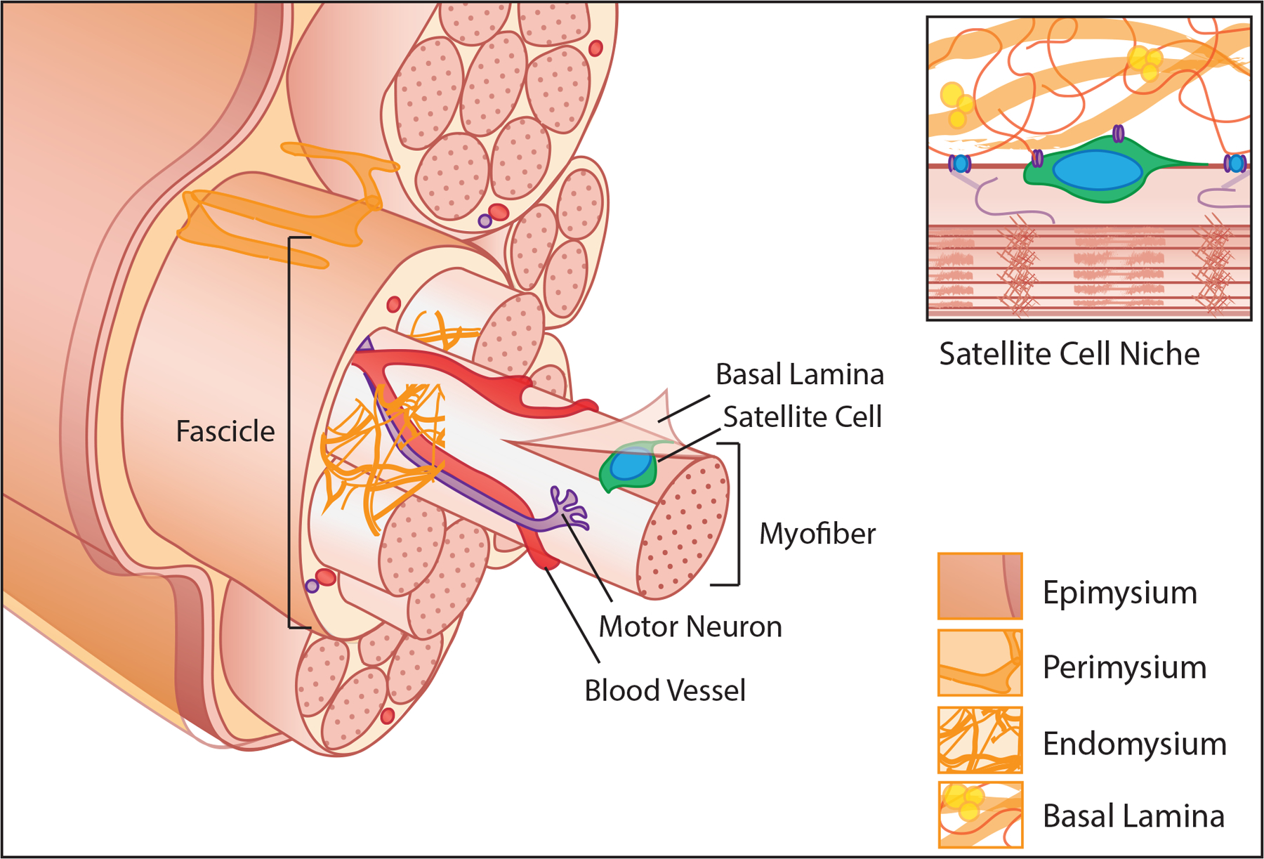
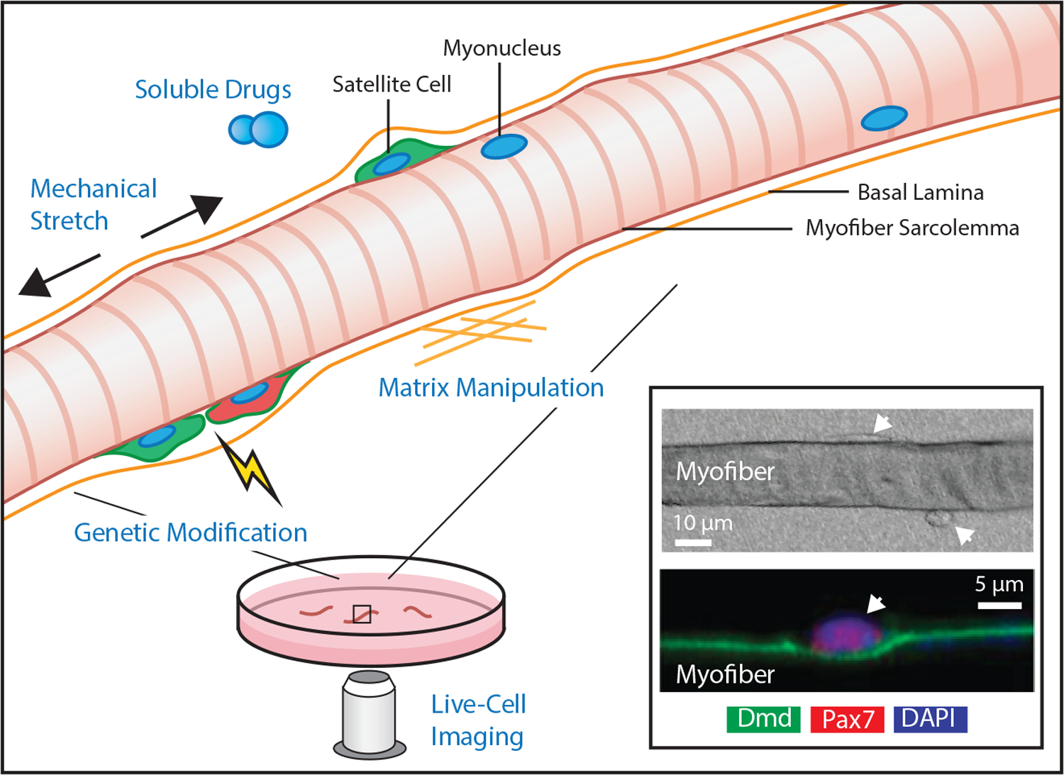
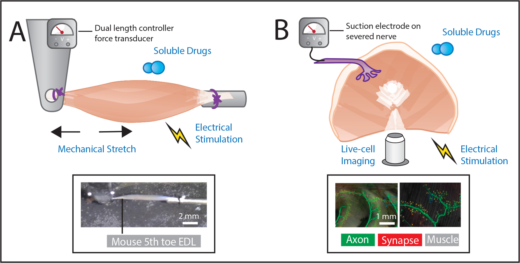
Purpose/Aim: Skeletal muscle tissue explants have been cultured and studied for nearly 100 years. These cultures, which retain complex tissue structure in an environment suited to precision manipulation and measurement, have led to seminal discoveries of the extrinsic and intrinsic mechanisms regulating contractility, metabolism and regeneration. This review discusses the two primary models of muscle explant: isolated myofiber and intact muscle.Materials and Methods: Relevant literature was reviewed and synthesized with a focus on the unique challenges and capabilities of each explant model.Results: Impactful past, current and future novel applications are discussed.Conclusions: Experiments using skeletal muscle explants have been integral to our understanding of the fundamentals of muscle physiology. As they are refined and adapted, they are poised to continue to inform the field for years to come.
Keywords: Isolated myofiber; ex-vivo stimulation; in-vitro culture; intact muscle; satellite cell.
Figures
Similar articles
-
Satellite cells and the muscle stem cell niche.Physiol Rev. 2013 Jan;93(1):23-67. doi: 10.1152/physrev.00043.2011. Physiol Rev. 2013. PMID: 23303905 Free PMC article. Review.
-
Regulatory factors and cell populations involved in skeletal muscle regeneration.J Cell Physiol. 2010 Jul;224(1):7-16. doi: 10.1002/jcp.22127. J Cell Physiol. 2010. PMID: 20232319 Review.
-
Satellite Cells and Skeletal Muscle Regeneration.Compr Physiol. 2015 Jul 1;5(3):1027-59. doi: 10.1002/cphy.c140068. Compr Physiol. 2015. PMID: 26140708 Review.
-
Isolation, Culture, and Immunostaining of Skeletal Muscle Myofibers from Wildtype and Nestin-GFP Mice as a Means to Analyze Satellite Cell.Methods Mol Biol. 2017;1556:51-102. doi: 10.1007/978-1-4939-6771-1_4. Methods Mol Biol. 2017. PMID: 28247345
-
Myogenic Satellite Cells: Biological Milieu and Possible Clinical Applications.Pak J Biol Sci. 2017;20(1):1-11. doi: 10.3923/pjbs.2017.1.11. Pak J Biol Sci. 2017. PMID: 29023009 Review.
Cited by
-
Bioprocessing Considerations towards the Manufacturing of Therapeutic Skeletal and Smooth Muscle Cells.Bioengineering (Basel). 2023 Sep 9;10(9):1067. doi: 10.3390/bioengineering10091067. Bioengineering (Basel). 2023. PMID: 37760170 Free PMC article. Review.
-
In vivo shear wave elasticity imaging for assessment of diaphragm function in muscular dystrophy.Acta Biomater. 2023 Sep 15;168:277-285. doi: 10.1016/j.actbio.2023.07.009. Epub 2023 Jul 13. Acta Biomater. 2023. PMID: 37453552 Free PMC article.
-
ERG1A K+ channel increases intracellular calcium concentration through modulation of calsequestrin1 in C2C12 myotubes.Sci Rep. 2025 Mar 19;15(1):9480. doi: 10.1038/s41598-025-93788-7. Sci Rep. 2025. PMID: 40108273 Free PMC article.
-
Experimental Models of Sarcopenia: Bridging Molecular Mechanism and Therapeutic Strategy.Cells. 2020 Jun 2;9(6):1385. doi: 10.3390/cells9061385. Cells. 2020. PMID: 32498474 Free PMC article. Review.
-
Generating intrafusal skeletal muscle fibres in vitro: Current state of the art and future challenges.J Tissue Eng. 2020 Dec 29;11:2041731420985205. doi: 10.1177/2041731420985205. eCollection 2020 Jan-Dec. J Tissue Eng. 2020. PMID: 34956586 Free PMC article. Review.
References
-
- Strangeways TSP, Fell HB. Experimental studies on the differentiation of embryonic tissues growing in vivo and in vitro.—II. The development of the isolated early embryonic eye of the fowl when cultivated in vitro. Proc. Roy. Soc. London B; 1926.
-
- Hill AV. The heat of shortening and the dynamic constants of muscle. Proceedings of the Royal Society B; 1938. p. 136:195. - PubMed
-
- Ramsey RW, Street SF. The isometric length‐tension diagram of isolated skeletal muscle fibers of the frog. Journal of Cellular and Comparative Physiology; 1940. p. 11:34.
Publication types
MeSH terms
Grants and funding
LinkOut - more resources
Full Text Sources
Other Literature Sources
Research Materials
Miscellaneous