

Ciliary exclusion of Polycystin-2 promotes kidney cystogenesis in an autosomal dominant polycystic kidney disease model
- PMID: 31492868
- PMCID: PMC6731238
- DOI: 10.1038/s41467-019-12067-y
Ciliary exclusion of Polycystin-2 promotes kidney cystogenesis in an autosomal dominant polycystic kidney disease model
Abstract
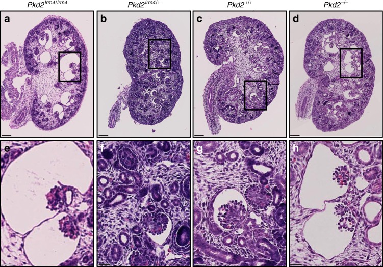
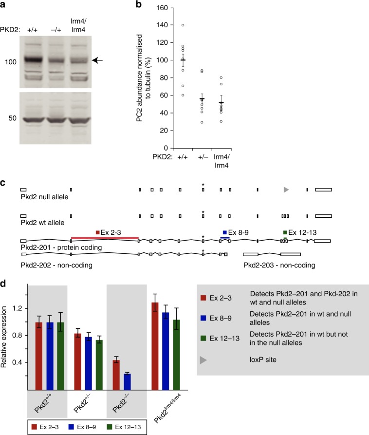
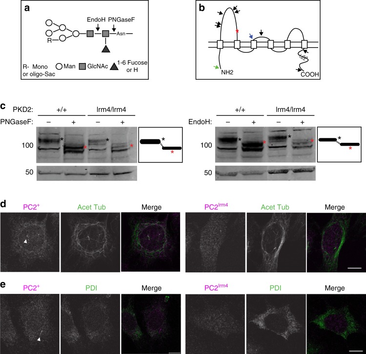
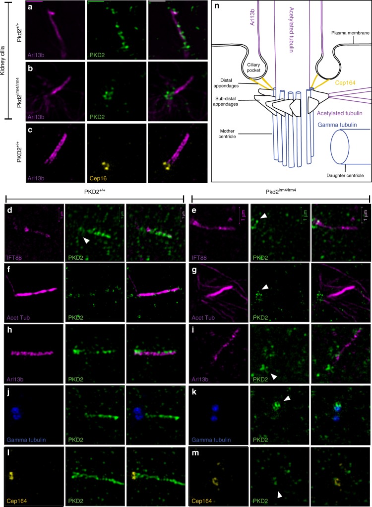
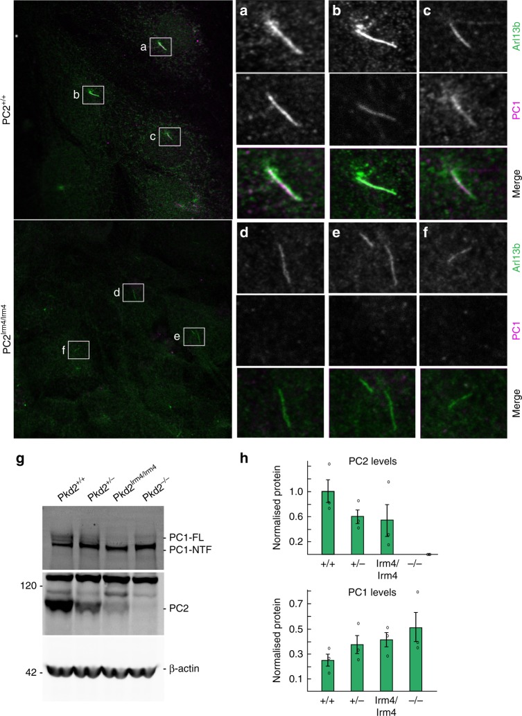
The human PKD2 locus encodes Polycystin-2 (PC2), a TRPP channel that localises to several distinct cellular compartments, including the cilium. PKD2 mutations cause Autosomal Dominant Polycystic Kidney Disease (ADPKD) and affect many cellular pathways. Data underlining the importance of ciliary PC2 localisation in preventing PKD are limited because PC2 function is ablated throughout the cell in existing model systems. Here, we dissect the ciliary role of PC2 by analysing mice carrying a non-ciliary localising, yet channel-functional, PC2 mutation. Mutants develop embryonic renal cysts that appear indistinguishable from mice completely lacking PC2. Despite not entering the cilium in mutant cells, mutant PC2 accumulates at the ciliary base, forming a ring pattern consistent with distal appendage localisation. This suggests a two-step model of ciliary entry; PC2 first traffics to the cilium base before TOP domain dependent entry. Our results suggest that PC2 localisation to the cilium is necessary to prevent PKD.
Conflict of interest statement
The authors declare no competing interests.
Figures
Similar articles
-
Regulation of polycystin expression, maturation and trafficking.Cell Signal. 2020 Aug;72:109630. doi: 10.1016/j.cellsig.2020.109630. Epub 2020 Apr 8. Cell Signal. 2020. PMID: 32275942 Free PMC article. Review.
-
Reduced ciliary polycystin-2 in induced pluripotent stem cells from polycystic kidney disease patients with PKD1 mutations.J Am Soc Nephrol. 2013 Oct;24(10):1571-86. doi: 10.1681/ASN.2012111089. Epub 2013 Sep 5. J Am Soc Nephrol. 2013. PMID: 24009235 Free PMC article.
-
Cilia and polycystic kidney disease.Semin Cell Dev Biol. 2021 Feb;110:139-148. doi: 10.1016/j.semcdb.2020.05.003. Epub 2020 May 28. Semin Cell Dev Biol. 2021. PMID: 32475690 Review.
-
Polycystin-2-dependent transcriptome reveals early response of autosomal dominant polycystic kidney disease.Physiol Genomics. 2023 Nov 1;55(11):565-577. doi: 10.1152/physiolgenomics.00040.2023. Epub 2023 Sep 18. Physiol Genomics. 2023. PMID: 37720991 Free PMC article.
-
Regulation of ciliary trafficking of polycystin-2 and the pathogenesis of autosomal dominant polycystic kidney disease.Zhong Nan Da Xue Xue Bao Yi Xue Ban. 2010 Feb;35(2):93-9. doi: 10.3969/j.issn.1672-7347.2010.02.001. Zhong Nan Da Xue Xue Bao Yi Xue Ban. 2010. PMID: 20197605 Review.
Cited by
-
Regulation of polycystin expression, maturation and trafficking.Cell Signal. 2020 Aug;72:109630. doi: 10.1016/j.cellsig.2020.109630. Epub 2020 Apr 8. Cell Signal. 2020. PMID: 32275942 Free PMC article. Review.
-
Insights Into the Molecular Mechanisms of Polycystic Kidney Diseases.Front Physiol. 2021 Sep 8;12:693130. doi: 10.3389/fphys.2021.693130. eCollection 2021. Front Physiol. 2021. PMID: 34566674 Free PMC article. Review.
-
Ciliary IFT88 Protects Coordinated Adolescent Growth Plate Ossification From Disruptive Physiological Mechanical Forces.J Bone Miner Res. 2022 Jun;37(6):1081-1096. doi: 10.1002/jbmr.4502. Epub 2022 Feb 20. J Bone Miner Res. 2022. PMID: 35038201 Free PMC article.
-
Molecular and structural perspectives on protein trafficking to the primary cilium membrane.Biochem Soc Trans. 2024 Jun 26;52(3):1473-1487. doi: 10.1042/BST20231403. Biochem Soc Trans. 2024. PMID: 38864436 Free PMC article. Review.
-
Disease-associated missense mutations in the pore loop of polycystin-2 alter its ion channel function in a heterologous expression system.J Biol Chem. 2024 Aug;300(8):107574. doi: 10.1016/j.jbc.2024.107574. Epub 2024 Jul 14. J Biol Chem. 2024. PMID: 39009345 Free PMC article.
References
Publication types
MeSH terms
Substances
Grants and funding
LinkOut - more resources
Full Text Sources
Other Literature Sources
Molecular Biology Databases
Miscellaneous
