. 2019 Sep 6;9(1):12854.
doi: 10.1038/s41598-019-49314-7.

Validation and assessment of an antibiotic-based, aseptic decontamination manufacturing protocol for therapeutic, vacuum-dried human amniotic membrane

Affiliations

Validation and assessment of an antibiotic-based, aseptic decontamination manufacturing protocol for therapeutic, vacuum-dried human amniotic membrane

Nagi M Marsit et al. Sci Rep. .

Abstract

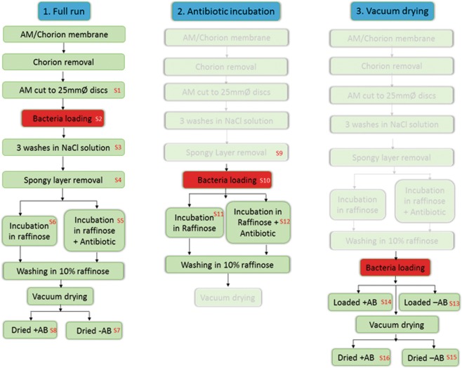
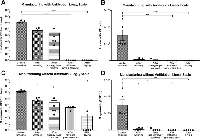
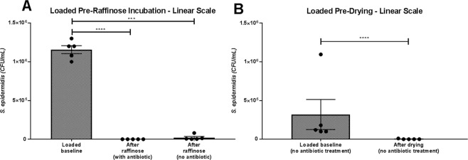
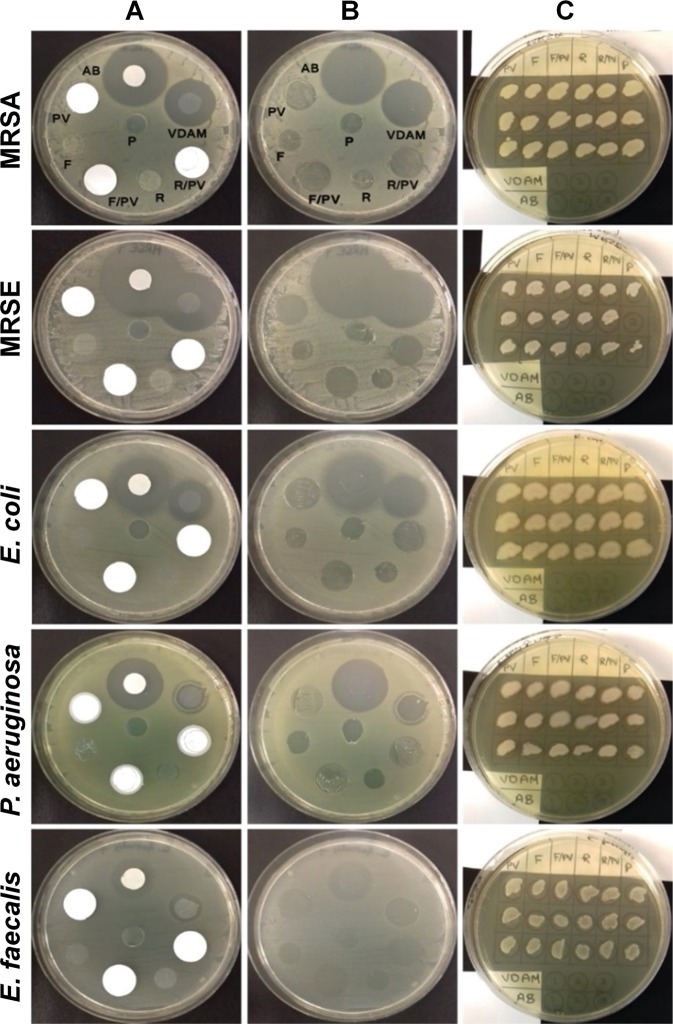
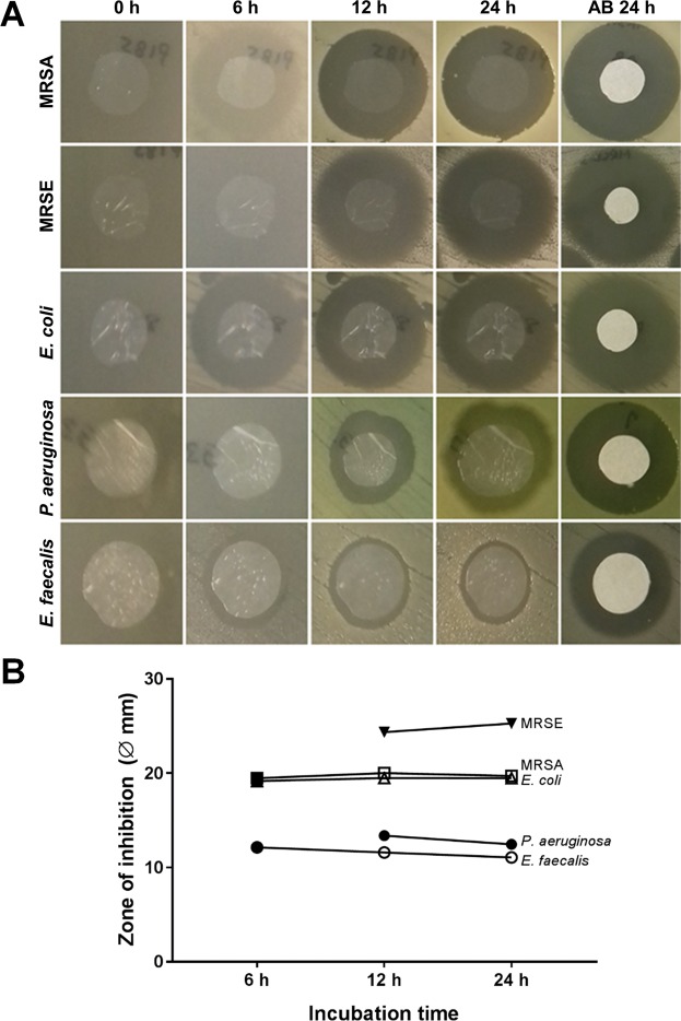
Amniotic membrane (AM) is used to treat a range of ophthalmic indications but must be presented in a non-contaminated state. AM from elective caesarean sections contains natural microbial contamination, requiring removal during processing protocols. The aim of this study was to assess the ability of antibiotic decontamination of AM, during processing by innovative low-temperature vacuum-drying. Bioburden of caesarean section AM was assessed, and found to be present in low levels. Subsequently, the process for producing vacuum-dried AM (VDAM) was assessed for decontamination ability, by artificially loading with Staphylococcus epidermidis at different stages of processing. The protocol was highly efficient at removing bioburden introduced at any stage of processing, with antibiotic treatment and drying the most efficacious steps. The antibacterial activity of non-antibiotic treated AM compared to VDAM was evaluated using minimum inhibitory/biocidal concentrations (MIC/MBC), and disc diffusion assays against Meticillin-resistant Staphylococcus aureus, Meticillin-resistant S. epidermidis, Escherichia coli, Pseudomonas aeruginosa and Enterococcus faecalis. Antibacterial activity without antibiotic was low, confirmed by high MIC/MBC, and a no inhibition on agar lawns. However, VDAM with antibiotic demonstrated effective antibacterial capacity against all bacteria. Therefore, antibiotic decontamination is a reliable method for sterilisation of AM and the resultant antibiotic reservoir is effective against gram-positive and -negative bacteria.


Conflict of interest statement

Dr. Hopkinson and Dr. Allen own shares in NuVision Biotherapies. Dr. Hopkinson, Dr. Britchford and Mr. McIntosh were partially employed by NuVision Biotherapies whilst this study took place. Dr. Sidney, Mr. Ashraf and Prof. Bayston declare no potential conflict of interest.

Figures

Figure 1
Figure 1
Schematic diagram showing sampling procedures for the S. epidermidis loaded bioburden experiment. Discs of 25 mm diameter were collected at each point numbered in red (S1–S15). Experiment was performed on 5 separate donors.
Figure 2
Figure 2
Effect of VDAM processing on reduction of artificially loaded S. epidermidis bacterial counts. Loading of 100 μL of 106 CFU/mL S. epidermidis was performed before washing procedures in 0.9% NaCl. AM samples were taken throughout processing and assessed for CFU/mL. Manufacturing was performed with antibiotic included in the raffinose step (A) graph shown with log10 scale, (B) graph shown with linear scale. Manufacturing with antibiotic included (C) graph shown with log10 scale, (D) graph shown with linear scale. Data shows individual experiments performed with 5 different donors and is represented as mean ± SEM. Data compared by one-way ANOVA, Kruskal-Wallis test followed by multiple comparison uncorrected Dunn’s test. Statistical significance vs. baseline represented by *p ≤ 0.05, ***p ≤ 0.001, ****p ≤ 0.0001.
Figure 3
Figure 3
Efficiency of antibiotic incubation and drying steps in reducing artificially loaded S. epidermidis bacterial counts. (A) Loading of 100 μL of 106S. epidermidis was performed before raffinose incubation with/without antibiotic. AM samples were taken after loading and after incubation and assessed for CFU/mL. (B) Loading of 100 μL of 106 CFU/mL S. epidermidis was performed after raffinose incubation with/without antibiotic and before drying. AM samples were taken after loading and after drying and assessed for CFU/mL. Only samples processed without antibiotic are shown, as no colonies were seen either in the loaded baseline or after drying. Data represents experiments performed on 5 different donors and is represented as mean ± SEM. Data compared by one-way ANOVA, Kruskal-Wallis test followed by multiple comparison uncorrected Dunn’s test. Statistical significance vs. baseline represented by ***p ≤ 0.001, ****p ≤ 0.0001.
Figure 4
Figure 4
Antibacterial activity of various amniotic membrane preparations. Discs of 10 mm diameter were prepared of VDAM; non-raffinose and non-antibiotic treated AM (F), non-antibiotic treated AM (R) and antibiotic soaked Whatman no. 3 filter paper (AB) as a positive control. Polyvinylidene fluoride (PVDF) transfer membrane discs (PV) were used as a control to the following disc samples: F over PVDF (F/PV) and R over PVDF (R/PV). The transparent plastic material (P) was used as a carrier to transfer VDAM onto new for a further incubation period. All discs were laid on lawns of MRSA, MRSE, E. coli, P. aeruginosa and E. faecalis. Images show representative MHA plates after overnight incubation at 35 °C. (A) Bacterial lawns with discs still present. (B) Same plates after removal of samples. (C) Streaks from areas underneath discs, shown in B, onto lawns of corresponding bacteria on MHA plates after 35 °C overnight incubation. Experimental error caused MRSE sample P2 not to be streaked. Experiment conducted on three different donors.
Figure 5
Figure 5
Antibiotic release from VDAM over time. (A) Antibiotic release from 10 mm diameter discs of VDAM placed on bacterial lawns. Representative images show zones of inhibition of the same disc after 6, 12 and 24 hours of incubation at 35 °C. Filter paper discs impregnated in antibiotic were used as control in every plate. Experiment performed on triplicate samples from 3 different donors and repeated 3 times for all bacteria. (B) Quantification of the zone of inhibition (including disc diameter) at the 6, 12 and 24 hour time points.

References

    1. Azuara-Blanco A, Pillai C, Dua HS. Amniotic membrane transplantation for ocular surface reconstruction. Br. J. Ophthalmol. 1999;83:399–402. doi: 10.1136/bjo.83.4.399. - DOI - PMC - PubMed
    1. Gicquel JJ, Bejjani RA, Ellies P, Mercie M, Dighiero P. Amniotic membrane transplantation in severe bacterial keratitis. Cornea. 2007;26:27–33. doi: 10.1097/ICO.0b013e31802b28df. - DOI - PubMed
    1. Liu J, Sheha H, Fu Y, Liang L, Tseng SCG. Update on amniotic membrane transplantation. Expert Rev. Ophthalmol. 2010;5:645–661. doi: 10.1586/eop.10.63. - DOI - PMC - PubMed
    1. Kim JS, Kim JC, Hahn TW, Park WC. Amniotic membrane transplantation in infectious corneal ulcer. Cornea. 2001;20:720–726. doi: 10.1097/00003226-200110000-00010. - DOI - PubMed
    1. Bouchard CS, John T. Amniotic membrane transplantation in the management of severe ocular surface disease: Indications and outcomes. Ocul. Surf. 2004;2:201–211. doi: 10.1016/S1542-0124(12)70062-9. - DOI - PubMed

Publication types