. 2019 Sep 6;9(1):12861.
doi: 10.1038/s41598-019-49339-y.

Exosomes from Adipose-Derived Stem Cells (ADSCs) Overexpressing miR-21 Promote Vascularization of Endothelial Cells

Affiliations

Exosomes from Adipose-Derived Stem Cells (ADSCs) Overexpressing miR-21 Promote Vascularization of Endothelial Cells

Yang An et al. Sci Rep. .

Abstract

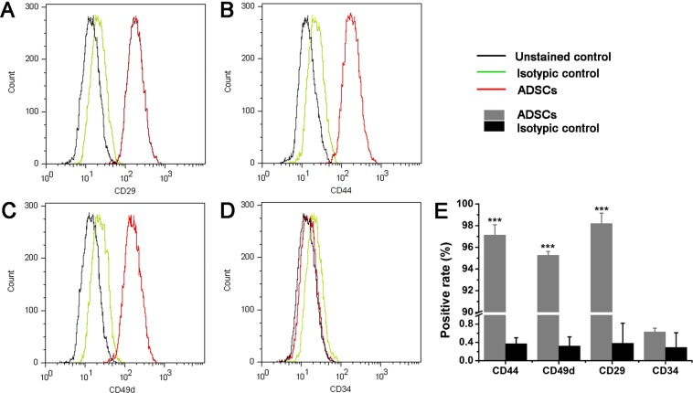
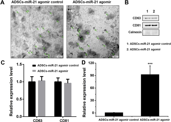
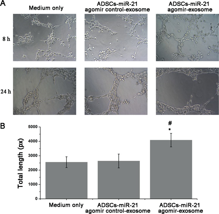
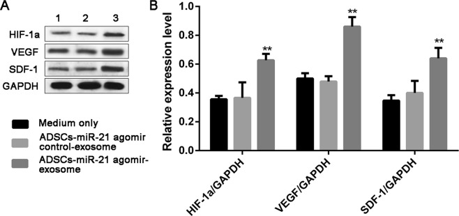
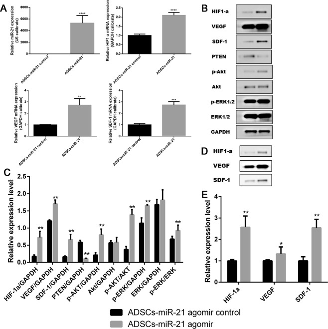
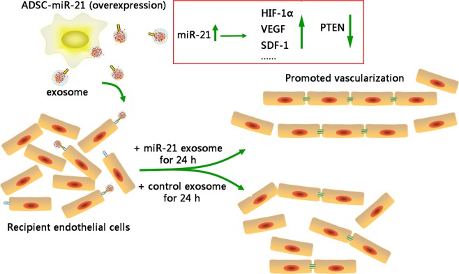
In the past few years, exosomes released from adipose-derived stem cells (abbreviated as ADSCs) have shown promises to provide therapeutic benefits in the fields of regenerative medicine. miRNAs, existing in exosomes, are endogenous, small noncoding RNAs that play important roles in a variety of cellular functions and tumor development. Emerging evidences have indicated that miR-21 is one of the important miRNAs associated with tumor angiogenesis. In this study, we identified the role of exosomes from ADSCs overexpressing miR-21 in regulating/promoting vascularization of endothelial cells. Experimental data indicated an elevated miR-21 level in exosomes released by ADSCs overexpressing miR-21. In vitro matrigel angiogenesis assay showed that exosomes secreted by ADSCs overexpressing miR-21 significantly promoted the vascularization of HUVEC cells (an endothelial cell line). Quantitative real-time polymerase chain reaction (qRT-PCR) and western blot (WB) revealed an upregulation of HIF-1α, VEGF, SDF-1, p-Akt, p-ERK1/2 and downregulation of PTEN in response to miR-21 overexpression, indicating that miR-21 enriched exosomes induced angiogenesis through Akt and ERK activation and also HIF-1α and SDF-1 expression. Our work suggests that exosomes from ADSCs that overexpressing miR-21 can potentially promote vascularization and therefore the transplantation of exosomes from their culture may be suitable for clinical effort in regenerative medicine.


Conflict of interest statement

The authors declare no competing interests.

Figures

Figure 1
Figure 1
Characterization of ADSCs isolated from inguinal fat pad of 3-week old Lewis male rats by flow cytometric analysis. (AD) Flow cytometric analysis of ADSC surface markers (CD29, CD44, CD49d and CD34). The results presented are typical of those obtained from three separate experiments. (E) Summary of the result in (AD). Error bars were calculated based on triplicates. (***means P < 0.001 vs. isotypic control).
Figure 2
Figure 2
The protocol for exosome extraction from ADSCs.
Figure 3
Figure 3
Characterization of exosomes derived from ADSCs isolated from inguinal fat pad of 3-week old Lewis male rats by flow cytometric analysis. (A) TEM images show the ultrastructure of ADSC-derived exosomes. Scale bar was 200 nm. (B) Expression of the exosome markers CD63, CD81, and Calnexin confirmed by western blot. (C) Quantification of the expression of the exosome markers CD63 and CD81 in (B). (D) The level of miR-21 in exosomes secreted from ADSCs with or without miR-21 overexpression. Error bars were calculated based on triplicates. (***means p < 0.001 vs. ADSCs- miR-21 agomir control).
Figure 4
Figure 4
Exosomes from ADSCs overexpressing miR-21 promote vascularization of endothelial cells (HUVEC). (A) Tube formation capability detected in endothelial cells (HUVEC) stimulated with 5 μg/ml ADSCs-miR-21 agomir control-exosome or ADSCs-miR-21 agomir exosome for 8 or 24 h. (B) Quantification of the length of the formed tubes at 8 h in (A). Error bars were calculated based on triplicates. (*means P < 0.05 vs. medium only; #means P < 0.05 vs. ADSCs- miR-21 agomir control-exosome).
Figure 5
Figure 5
Exosomes from ADSCs overexpressing miR-21 enhanced expression of HIF-1α, VEGF and SDF-1 in HUVEC. Western blot (A) and its quantification (B) of the expression of HIF-1α, VEGF, SDF-1 in HUVEC with control medium (1), exosomes from control ADSCs (2) and exosomes from miR-21 overexpressing ADSCs. Error bars were calculated based on triplicates. (**Means p < 0.01 vs. both medium only and ADSCs- miR-21 agomir control-exosome).
Figure 6
Figure 6
Overexpression of miR-21 in ADSCs leads to the upregulation of HIF-1α, VEGF, SDF-1, p-Akt, p-ERK1/2 and downregulation of PTEN. (A) Quantification of the expression level of miR-21, HIF-1α, VEGF, SDF-1 in ADSCs with and without miR-21 overexpression. (B,C) Western blot and its quantification of the expression level of HIF-1α, VEGF, SDF-1, PTEN, p-Akt, Akt, p-ERK1/2, ERK1/2 in ADSCs with (2) and without miR-21 overexpression (1). (D,E) Western blot and its quantification of the expression level of HIF-1α, VEGF, SDF-1 in exosomes isolated from ADSCs with (2) and without (1) miR-21 overexpression. Error bars were calculated based on triplicates. (***means p < 0.001 vs. ADSCs- miR-21 control, **means p < 0.01 vs. ADSCs- miR-21 control, *means p < 0.05 vs. ADSCs- miR-21 control).
Figure 7
Figure 7
The graphic summary of miR-21 overexpressing exosome promoting vascularization of endothelial cells.

Similar articles

Cited by

References

    1. Skog J, et al. Glioblastoma microvesicles transport RNA and proteins that promote tumour growth and provide diagnostic biomarkers. Nature Cell Biology. 2008;10:1470–U1209. doi: 10.1038/ncb1800. - DOI - PMC - PubMed
    1. Thery C, Zitvogel L, Amigorena S. Exosomes: Composition, biogenesis and function. Nature Reviews Immunology. 2002;2:569–579. doi: 10.1038/nri855. - DOI - PubMed
    1. Valadi H, et al. Exosome-mediated transfer of mRNAs and microRNAs is a novel mechanism of genetic exchange between cells. Nature Cell Biology. 2007;9:654–U672. doi: 10.1038/ncb1596. - DOI - PubMed
    1. Oksvold MP, et al. Expression of B-Cell Surface Antigens in Subpopulations of Exosomes Released From B-Cell Lymphoma Cells. Clinical Therapeutics. 2014;36:847–862. doi: 10.1016/j.clinthera.2014.05.010. - DOI - PubMed
    1. Gruenberg J. The endocytic pathway: A mosaic of domains. Nat. Rev. Mol. Cell Biol. 2001;2:721–730. doi: 10.1038/35096054. - DOI - PubMed

Publication types

MeSH terms

Substances