

Solochrome cyanine: A histological stain for cobalt-chromium wear particles in metal-on-metal periprosthetic tissues
- PMID: 31493091
- PMCID: PMC6731196
- DOI: 10.1007/s10856-019-6304-0
Solochrome cyanine: A histological stain for cobalt-chromium wear particles in metal-on-metal periprosthetic tissues
Erratum in
-
Correction to: Solochrome cyanine: A histological stain for cobalt-chromium wear particles in metal-on-metal periprosthetic tissues.J Mater Sci Mater Med. 2019 Sep 25;30(9):110. doi: 10.1007/s10856-019-6312-0. J Mater Sci Mater Med. 2019. PMID: 31555914 Free PMC article.
Abstract
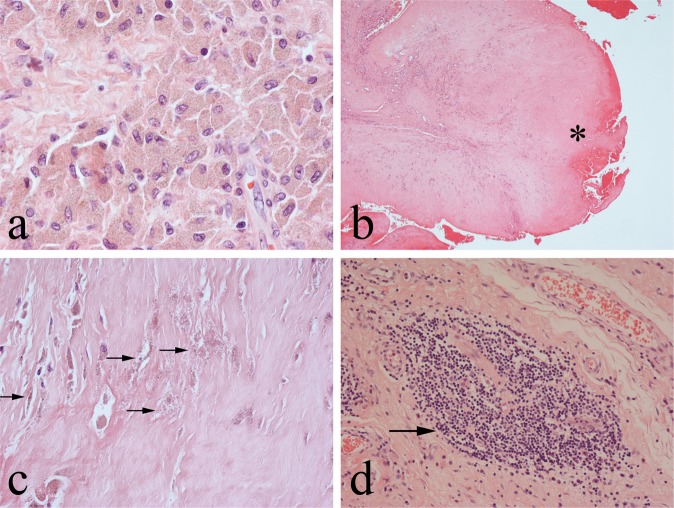
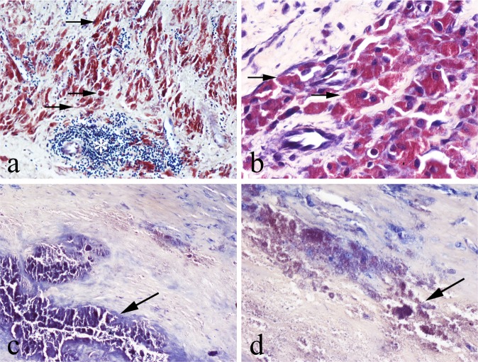
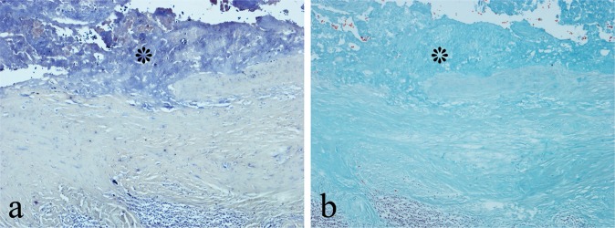
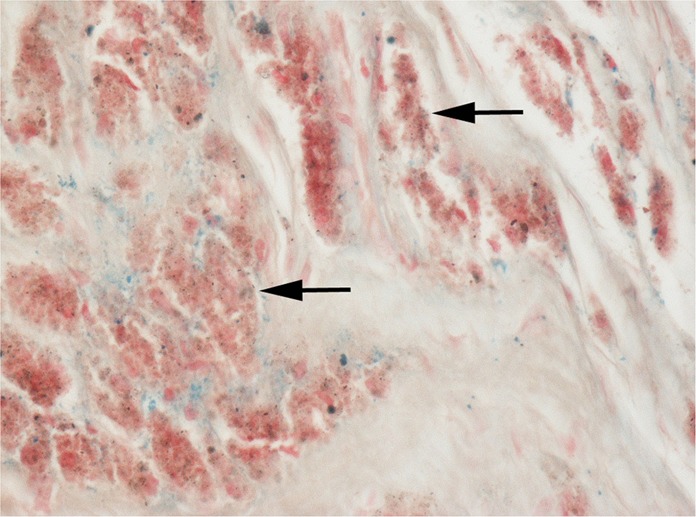
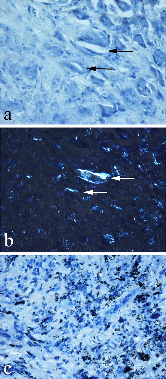
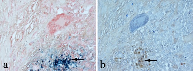
Metal-on-metal (MoM) hip arthroplasties produce abundant implant-derived wear debris composed mainly of cobalt (Co) and chromium (Cr). Cobalt-chromium (Co-Cr) wear particles are difficult to identify histologically and need to be distinguished from other wear particle types and endogenous components (e.g., haemosiderin, fibrin) which may be present in MoM periprosthetic tissues. In this study we sought to determine whether histological stains that have an affinity for metals are useful in identifying Co-Cr wear debris in MoM periprosthetic tissues. Histological sections of periprosthetic tissue from 30 failed MoM hip arthroplasties were stained with haematoxylin-eosin (HE), Solochrome Cyanine (SC), Solochrome Azurine (SA) and Perls' Prussian Blue (PB). Sections of periprosthetic tissue from 10 cases of non-MoM arthroplasties using other implant biomaterials, including titanium, ceramic, polymethylmethacrylate (PMMA) and ultra-high molecular weight polyethylene (UHMWP) were similarly analysed. Sections of 10 cases of haemosiderin-containing knee tenosynovial giant cell tumour (TSGCT) were also stained with HE, SC, SA and PB. In MoM periprosthetic tissues, SC stained metal debris in phagocytic macrophages and in the superficial necrotic zone which exhibited little or no trichrome staining for fibrin. In non-MoM periprosthetic tissues, UHMWP, PMMA, ceramic and titanium particles were not stained by SC. Prussian Blue, but not SC or SA, stained haemosiderin deposits in MoM periprosthetic tissues and TSGT. Our findings show that SC staining (most likely Cr-associated) is useful in distinguishing Co-Cr wear particles from other metal/non-metal wear particles types in histological preparations of periprosthetic tissue and that SC reliably distinguishes haemosiderin from Co-Cr wear debris.
Conflict of interest statement
NAA and HSG have provided medicolegal opinions in arthroplasty-related cases. The other authors declare that they have no conflict of interest.
Figures
Similar articles
-
Periprosthetic tissue metal content but not serum metal content predicts the type of tissue response in failed small-diameter metal-on-metal total hip arthroplasties.J Bone Joint Surg Am. 2013 Sep 4;95(17):1561-8. doi: 10.2106/JBJS.L.01273. J Bone Joint Surg Am. 2013. PMID: 24005196
-
Analysis of bearing wear, whole blood and synovial fluid metal ion concentrations and histopathological findings in patients with failed ASR hip resurfacings.BMC Musculoskelet Disord. 2017 Dec 11;18(1):523. doi: 10.1186/s12891-017-1894-5. BMC Musculoskelet Disord. 2017. PMID: 29228956 Free PMC article.
-
Association between periprosthetic tissue metal content, whole blood and synovial fluid metal ion levels and histopathological findings in patients with failed metal-on-metal hip replacement.PLoS One. 2018 May 16;13(5):e0197614. doi: 10.1371/journal.pone.0197614. eCollection 2018. PLoS One. 2018. PMID: 29768492 Free PMC article.
-
Understanding outcomes and toxicological aspects of second generation metal-on-metal hip implants: a state-of-the-art review.Crit Rev Toxicol. 2018 Nov;48(10):853-901. doi: 10.1080/10408444.2018.1563048. Epub 2019 Mar 26. Crit Rev Toxicol. 2018. PMID: 30912993 Review.
-
Can cobalt levels estimate in-vivo wear of metal-on-metal bearings used in hip arthroplasty?Proc Inst Mech Eng H. 2007 Nov;221(8):929-42. doi: 10.1243/09544119JEIM270. Proc Inst Mech Eng H. 2007. PMID: 18161253 Review.
References
MeSH terms
Substances
LinkOut - more resources
Full Text Sources
