. 2019 Sep 7:25:6719-6726.
doi: 10.12659/MSM.915532.

Effects of Left Gastric Artery Ligation Versus Sleeve Gastrectomy on Obesity-Induced Adipose Tissue Macrophage Infiltration and Inflammation in Diet-Induced Obese Rats

Affiliations

Effects of Left Gastric Artery Ligation Versus Sleeve Gastrectomy on Obesity-Induced Adipose Tissue Macrophage Infiltration and Inflammation in Diet-Induced Obese Rats

Yi Shen et al. Med Sci Monit. .

Abstract

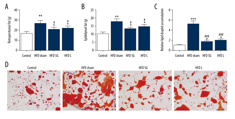
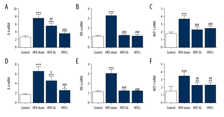
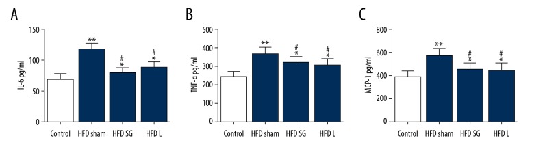
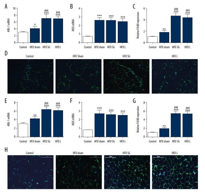
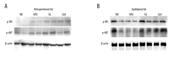
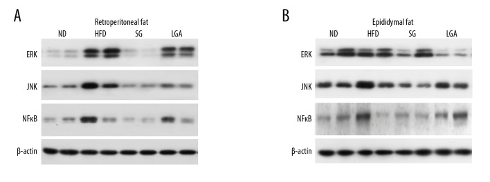
BACKGROUND Bariatric procedures such as left gastric artery ligation (LGAL) and sleeve gastrectomy (SG) have emerged as important procedures for treating morbid obesity. In this study, we compared the effects of LGAL vs. SG on obesity-induced adipose tissue macrophage infiltration and inflammation in diet-induced obese rats. MATERIAL AND METHODS Sprague-Dawley (SD) rats were fed a high-fat diet (HFD) for 16 weeks to induce obesity. SG, GLAL, or corresponding sham surgeries were performed in anesthetized rats. Inflammatory factor expression in serum and epididymal and retroperitoneal adipose tissues were analyzed 4 weeks after surgery. Macrophage infiltration and phenotype transformation were also assessed with Western blot analysis and immunofluorescence. RESULTS Both LGAL and SG strongly attenuated high-fat diet (HFD)-induced fat accumulation in retroperitoneal and epididymal tissues. The expressions of inflammatory cytokines such as tumor necrosis factor (TNF)-alpha, interleukin (IL)-6, and monocyte chemoattractant protein (MCP)-1 were downregulated after LGAL and after SG by promoting activation of M2 macrophages, despite continued exposure to HFD. Furthermore, both LGAL and SG resulted in increased macrophage infiltration, but did not contribute to phenotype transformation of macrophages to M1. CONCLUSIONS LGAL and SG both reduced fat accumulation caused by HFD feeding. Therapies designed to ameliorate the inflammatory response by promoting activation of M2 macrophages may be valuable.


Conflict of interest statement

Conflicts of interest

None.

Figures

Figure 1
Figure 1
Surgical treatment reversed high-fat diet (HFD)-induced fat accumulation of retroperitoneal and epididymal fat. Rats were fed with an HFD for 18 weeks before treatment with gastric left artery ligation and sleeve gastrectomy for 4 weeks. Whole adipose tissue from the retroperitoneum and epididymis were separated and weighed (A, B). Weight of whole adipose tissue from the retroperitoneum (A) and epididymis (B). Data are presented as the mean ±SD. * p<0.05, ** p<0.01 versus controls. # p<0.05 versus the HFD group. (C, D) Oil red staining shows lipid droplet deposition in fat sections.
Figure 2
Figure 2
The expression of inflammatory factors in retroperitoneal and epididymal adipose tissue. Rats were fed with an HFD for 18 weeks before gastric left artery ligation and sleeve gastrectomy for 4 weeks. Adipose tissue from the retroperitoneum and epididymis were stripped for Rt-PCR detection. (A–C) Expression level of IL-6 (A), TNF-α (B), and MCP-1 (C) from retroperitoneal fat in different groups. Data are presented as the mean ± SD. *** p<0.001 versus controls. ## p<0.01, ### p<0.001, HFD group. (D, E) The expression level of IL-6 (D), TNF-α (E), and MCP-1 (F) from epididymal fat in different groups. Data are presented as the mean ±SD. * p<0.05, ** p<0.01, *** p<0.001 versus the control. # p<0.05, ## p<0.01, ### p<0.001 versus the HFD group.
Figure 3
Figure 3
Inflammatory factor. (A–C) Serum inflammatory factor IL-6 (A), TNF-α (B) and MCP-1 in protein level were detection with ELISA. Data are presented as the mean ±SD. * p<0.05, ** p<0.01 versus the control. # p<0.05 versus the HFD group.
Figure 4
Figure 4
Macrophage infiltration and polarity conversion were detected with RT -PCR and immunofluorescence. (A, B) The expression level of M2 macrophage marker ARG-1 (A) and M1 macrophage marker iNOS (B) in adipose tissue from the retroperitoneum were detected with RT-PCR. Data are presented as the mean ±SD. * p<0.05, *** p<0.001 versus controls. ### p<0.001 versus the HFD group. (C, D) Macrophage infiltration in adipose (green) tissue from retroperitoneal adipose tissues were detected by immunofluorescence with F4/80 and DAPI (blue) staining. (E, F) The expression level of M2 macrophage marker ARG-1 (E) and M1 macrophage marker iNOS (F) in epididymal adipose tissue were detected with RT -PCR. Data are presented as the mean ±SD. ** p<0.01, *** p<0.001 versus controls. ### p<0.001 versus HFD group. (G, H) Macrophage infiltration in adipose (green) tissue from epididymal tissue was detected by immunofluorescence staining with F4/80 and DAPI (blue) staining.
Figure 5
Figure 5
The effect of gastric left artery ligation and sleeve gastrectomy on p-IRS-1 and p-Akt protein expression. (A) Western blot detection shows the expression of p-IRS-1 and p-Akt in adipose tissue from the retroperitoneal tissue. (B) Western blot detection shows the expression of p-IRS-1 and p-Akt in epididymal adipose tissue.
Figure 6
Figure 6
The effect of gastric left artery ligation and sleeve gastrectomy on inflammation and insulin signaling relative protein expression. (A) Western blot shows the expression of ERK, JNK, and NFκB in retroperitoneal adipose tissue. (B) Western blot detection shows the expression of ERK, JNK, and NF-κB in epididymal adipose tissue.

References

    1. Hotamisligil GS, Shargill NS, Spiegelman BM. Adipose expression of tumor necrosis factor-alpha: Direct role in obesity-linked insulin resistance. Science. 1993;259:87–91. - PubMed
    1. Xu H, Barnes GT, Yang Q, et al. Chronic inflammation in fat plays a crucial role in the development of obesity-related insulin resistance. J Clin Invest. 2003;112:1821–30. - PMC - PubMed
    1. Mauer J, Chaurasia B, Plum L, et al. Myeloid cell-restricted insulin receptor deficiency protects against obesity-induced inflammation and systemic insulin resistance. PLoS Genet. 2010;6:e1000938. - PMC - PubMed
    1. Weisberg SP, McCann D, Desai M, et al. Obesity is associated with macrophage accumulation in adipose tissue. J Clin Invest. 2003;112:1796–808. - PMC - PubMed
    1. Palmer JG. Liability considerations presented by human gene therapy. Hum Gene Ther. 1991;2:235–42. - PubMed