. 2019 Nov;23(11):7490-7504.
doi: 10.1111/jcmm.14617. Epub 2019 Sep 7.

The N-terminal D1 domain of Treponema pallidum flagellin binding to TLR5 is required but not sufficient in activation of TLR5

Affiliations

The N-terminal D1 domain of Treponema pallidum flagellin binding to TLR5 is required but not sufficient in activation of TLR5

Man Xu et al. J Cell Mol Med. 2019 Nov.

Abstract

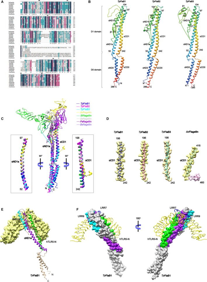
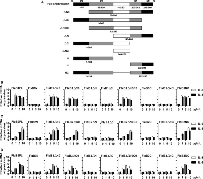
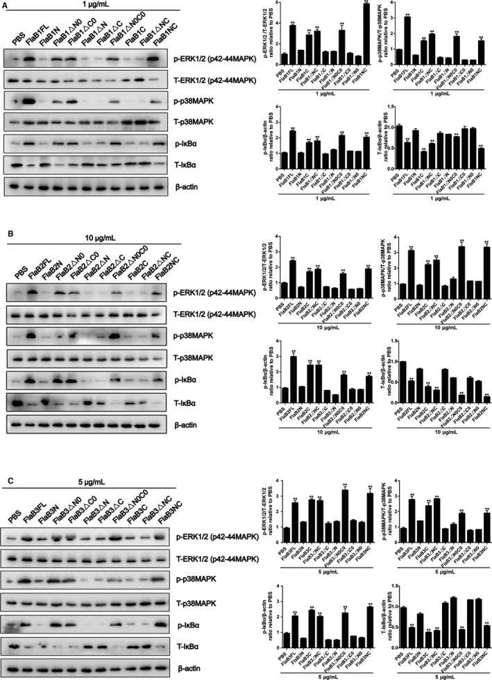
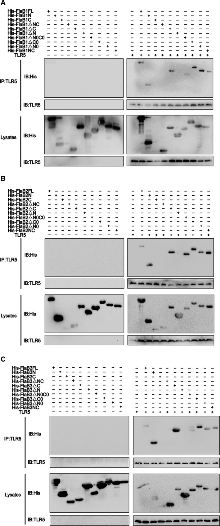
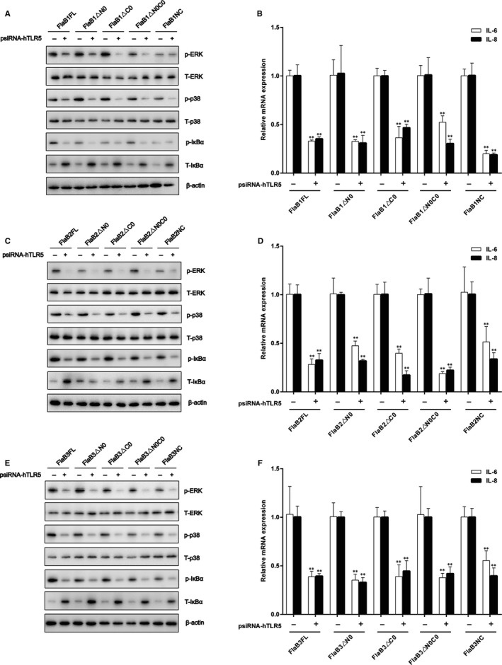
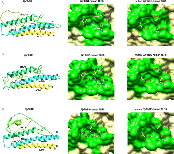
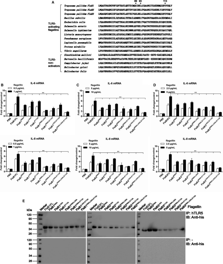
Syphilis is a chronic bacterial infection caused by Treponema pallidum (T pallidum) and the pathogenesis that T pallidum infection induces immunopathological damages in skin and other tissues remains unclear. We have previously reported that recombinant flagellins of T pallidum can elicit IL-6 and IL-8 transcriptions via TLR5 pathway. To identify the domains which induced the pro-inflammatory activity and the importance of the interactions between TLR5 and domains, homology-based modelling and comparative structural analyses revealed that Tpflagellins can combine with TLR5 directly. Deletion mutations showed that the ND1 domain binding to TLR5 is required but not sufficient in TLR5 activation. Moreover, site-directed mutagenesis analysis indicated that the arginine residue (Tpflagellins R89) of the ND1 domain and its adjacent residues (Tpflagellins L93 and E113) constitute a hot spot that elicits IL-6, IL-8 transcriptions and TLR5 activation, and affects the binding of Tpflagellins to TLR5. Taken together, these results give insight into the pathogenesis of T pallidum and may contribute to the future design of Tpflagellins-based therapeutics and syphilis vaccine.

Keywords: Treponema pallidum; N-terminal D1 domain; TLR5 signalling; flagellin; inflammation.


Conflict of interest statement

The authors declare no conflict of interest.

Figures

Figure 1
Figure 1
Modelling of Tpflagellins structures and structural comparison of various flagellins. A, Alignments of protein sequence from various flagellins. Protein sequences were retrieved from the Swiss‐Prot Database and aligned by ClustalW. Shading indicates areas where there is high conservation (dark) and moderate conservation (magenta and blue). Bsflagellin, Bacillus subtilis flagellin; Ecflagellin, Escherichia coli flagellin; Paflagellin, Pseudomonas aeruginosa; Stflagellin, Salmonella typhimurium flagellin; Sdflagellin, Salmonella enterica subspecies enterica serovar Dublin flagellin. B, Homology‐based structural model of Tpflagellins. The Tpflagellins structures are shown in rainbow‐coloured ribbons. Secondary structural elements are indicated. N, N‐terminus; C, C‐terminus. C, Overlays of the TpFlaB1 structure (blue ribbon), TpFlaB2 structure (magenta ribbon), TpFlaB3 structure (cyan ribbon) and other flagellin structures (coils) are shown [Sdflagellin (PDB ID 3V47, yellow), Stflagellin (PDB ID 1UCU, green), Ssflagellin (PDB ID 2ZBI, light grey), Paflagellin (PDB ID 4NX9, purple) and Bsflagellin (PDB ID 5GY2, cornflower blue)]. The αND1a and αCD1 regions of Tpflagellins adopted different conformations from various flagellins and are shown in ribbons in the left and right boxes, respectively. D, The αCD1 helices of Tpflagellins and Sdflagellin are shown as surface presentation and coils. E, Interactions of the TpFlaB1 with the hTLR5 chain. TpFlaB1 is shown in ribbons and hTLR5 chain is shown with yellow surface. F, Interactions of the TpFlaB1 with the hTLR5 chain. TpFlaB1 residues involved in hTLR5 binding are coloured by purple (αND1a), cyan (αND1b) and green (αCD1), hTLR5 chain is shown as yellow lines. The TLR5‐specific loops at LRR7 and LRR9 are shown as blue lines
Figure 2
Figure 2
The deletion mutant flagellins induce pro‐inflammatory mediators. A, Wild‐type and deletion mutant forms of Tpflagellins are represented schematically. Wild‐type Treponema pallidum protein consists of 286 amino acids and three functional domains. Amino acids 1‐139 represents N‐terminus, 202‐286 represents C‐terminus, and 140‐201 represents hypervariable region of flagellins. THP‐1 cells were stimulated with 1‐10 μg/mL FlaB1 (B) [FlaB2 (C) or FlaB3 (D)] mutant flagellins for 24 h. The gene transcription levels of IL‐6 and IL‐8 were analysed by qRT‐PCR. Data are presented as mean ± SD of three independent experiments
Figure 3
Figure 3
Mutant flagellins containing only D1 domains activate MAPK and NF‐κB. THP‐1 cells were treated with 1 μg/mL FlaB1 mutant flagellins (A), 10 μg/mL FlaB2 mutant flagellins (B) or 5 μg/mL FlaB3 mutant flagellins (C) for 1 h, and then, the total protein was extracted. The expression of nonphosphorylated and phosphorylated forms of ERK1/2 (p42‐44MAPK), p38MAPK and IκBα were analysed by Western blotting. ERK1/2 (p42‐44MAPK), p38MAPK and IκBα phosphorylation were quantified by ImageJ software and normalized with total protein or β‐actin. Data are presented as mean ± SD of three independent experiments. **P < .01 versus PBS control group
Figure 4
Figure 4
Mutant flagellins containing only ND1 domains bind to human TLR5. (A‐C) Co‐immunoprecipitation was performed to assess the binding of the mutated proteins to human TLR5. 30 μg of each protein was added to lysed THP‐1 cells (or lysis), then incubated with anti‐hTLR5 antibody at 4°C for 16 h, half of the lysates were incubated with 40 μL of beads for 1 h at room temperature and washed 5 times with wash buffer, and then, they were eluted and boiled for Western blotting analysis; the other half of the lysates were boiled and separated by SDS‐PAGE for Western blotting analysis. TLR5 polyclonal antibody was used for the detection of TLR5. Anti‐His antibodies were used for the detection of wild‐type flagellin and mutants
Figure 5
Figure 5
Identification of the D1 domain of Tpflagellins that affect TLR5 signalling. THP‐1 cells were transfected with psiRNA‐hTLR5 (psiRNA‐LucGL3 was used as a control) for 28 h, then stimulated with 1 μg/mL FlaB1 mutant flagellins (A), 10 μg/mL FlaB2 mutant flagellins (B), or 5 μg/mL FlaB3 mutant flagellins (C) for 1 h to analyse the expressions of nonphosphorylated and phosphorylated forms of ERK1/2 (p42‐44MAPK), p38MAPK, and IκBα, stimulated with 1 μg/mL FlaB1 mutant flagellins (D), 10 μg/mL FlaB2 mutant flagellins (E), or 5 μg/mL FlaB3 mutant flagellins (F) for 24 h to measure the transcription levels of IL‐6 and IL‐8 mRNA in the cells by using qRT‐PCR. Data are presented as mean ± SD of three independent experiments. *P < .05, **P < .01 versus corresponding control
Figure 6
Figure 6
Computational model structures of wild‐type Tpflagellins and Tpflagellins mutants. Residues Arg89, Leu93 and Glu113 of TpFlaB1 (A), TpFlaB2 (B) or TpFlaB3 (C) selected for alanine mutagenesis are present in wild‐type Tpflagellins structures (left). Residues Arg89, Leu93 and Glu113 of TpFlaB1 (A), TpFlaB2 (B) or TpFlaB3 (C) observed in the cavity formed by the human TLR5 LRR9 loop (green surface) in the Tpflagellin‐human TLR5 model (middle). Residues Alanine 89, Alanine 93 and Alanine 113 of TpFlaB1 (A), TpFlaB2 (B) or TpFlaB3 (C) observed in the cavity formed by the human TLR5 LRR9 loop (green surface) in the Tpflagellin‐human TLR5 model (right)
Figure 7
Figure 7
Identification of residues in the N‐terminal D1 domain of Tpflagellins that affect inflammation response. A, Amino acid alignment of ND1 domain flagellin sequences of several bacterial species and amino acid residues selected for alanine mutagenesis. B‐D, THP‐1 cells were stimulated with 0.5‐1 μg/mL FlaB1 mutant flagellins (B), 5‐10 μg/mL FlaB2 mutant flagellins (C) or 2.5‐5 μg/mL FlaB3 mutant flagellins (D) for 24 h. The gene transcription levels of IL‐6 and IL‐8 were analysed by qRT‐PCR. Data are presented as mean ± SD of three independent experiments. *P < .05, **P < .01 as compared with wild‐type flagellins. E, Co‐immunoprecipitation was performed to assess the binding of the mutated proteins to human TLR5. 30 μg of each protein was added to lysed THP‐1 cells (top) or control lysis (bottom) and then incubated with anti‐hTLR5 antibody at 4°C for 16 h, and the complexes were then incubated with 40 μL of beads for 1 h at room temperature and washed 5 times with wash buffer and eluted for Western blotting analysis. Anti‐His antibodies were used for the detection of wild‐type flagellin and mutants

Similar articles

Cited by

References

    1. Newman L, Rowley J, Vander Hoorn S, et al. Global estimates of the prevalence and incidence of four curable sexually transmitted infections in 2012 based on systematic review and global reporting. PLoS ONE. 2015;10:e0143304. - PMC - PubMed
    1. Ho EL, Lukehart SA. Syphilis: using modern approaches to understand an old disease. J Clin Investig. 2011;121:4584‐4592. - PMC - PubMed
    1. Wang C, Cheng Y, Liu B, et al. Sensitive detection of Treponema pallidum DNA from the whole blood of patients with syphilis by the nested PCR assay. Emerg Microbes Infect. 2018;7:83. - PMC - PubMed
    1. Edmondson DG, Hu B, Norris SJ. Long‐Term In Vitro Culture of the Syphilis Spirochete Treponema pallidum subsp. pallidum. mBio. 2018;7:9. - PMC - PubMed
    1. Luo X, Zhang X, Gan L, et al. The outer membrane protein Tp92 of Treponema pallidum induces human mononuclear cell death and IL‐8 secretion. J Cell Mol Med. 2018;22:6039‐6054. - PMC - PubMed

Publication types