. 2019 Dec 1:586:113414.
doi: 10.1016/j.ab.2019.113414. Epub 2019 Sep 4.

Efficient data acquisition with three-channel centerpieces in sedimentation velocity

Affiliations

Efficient data acquisition with three-channel centerpieces in sedimentation velocity

Kristian Juul-Madsen et al. Anal Biochem. .

Abstract

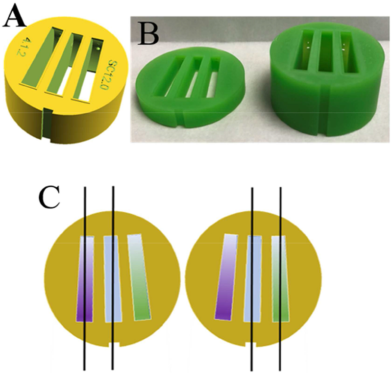
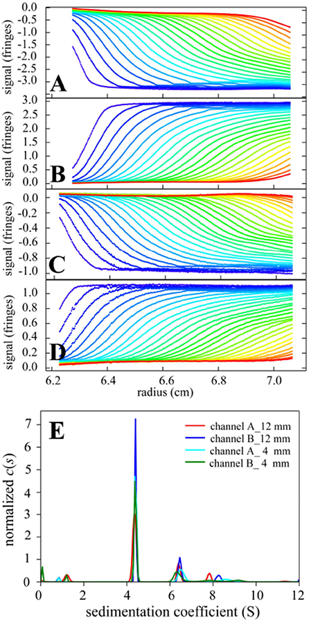
Three-channel 3D printed centerpieces with two sample sectors next to a joint solvent reference sector were recently described as a strategy to double the throughput of sedimentation velocity analytical ultracentrifugation experiments [Anal. Chem. 91 (2019) 5866-5873]. They are compatible with Rayleigh interference optical detection in commercial analytical ultracentrifuges, but require the rotor angles of data acquisition to be repeatedly adjusted during the experiment to record data from the two sample sectors. Here we present an approach to automate this data acquisition mode through the use of a secondary, general-purpose automation software, and an accompanying data pre-processing software for scan sorting.

Keywords: 3D printing; Analytical ultracentrifugation; Laboratory automation; Sedimentation velocity.


Figures

Figure 1.
Figure 1.
(A) Design of 3D printed three-channel centerpiece with sector angles of 2.2°, filling and venting holes, dome-shaped ceilings, and embossed rims surrounding the sectors that serve as integrated gaskets for vacuum seal. (B) Photographs of centerpieces 3D printed in an epoxy-like photopolymer “MicroFine Green” (ProtoLabs, Maple Plain, MN) using modification of a design file 3DPX-010758 of the NIH 3D Print Exchange (3dprint.nih.gov). Shown are centerpieces with 4 mm (left) and 12 mm pathlength (right), respectively. (C) Principle of illumination modes of three-channel centerpieces with Rayleigh interference optical detection. Two coherent parallel planar beams traverse either the left and the middle sector (left sketch), or the right and the middle sector (right sketch). The middle sector contains a joint reference buffer (blue), such that in the left configuration the purple sample can be observed, and in the right configuration the green sample can be observed. The two configurations have a 3-4° different rotation angle at the time of data acquisition, and due to the reversed beams they differ in the sign of the fringe shift signal.
Figure 2.
Figure 2.
(A-D) Sedimentation boundaries recorded in the left (A, C) and right (B, D) sample sectors of two three-channel centerpieces with 12 mm (A and B) and 4 mm (C and D) pathlength, respectively. (The 4 mm centerpiece was centered in height relative to the filling port, which allows measurement of gradients of up to ~100 fringes/cm [48].) Both sample sectors were filled with 1 mg/ml bovine serum albumin in phosphate buffered saline, with the buffer filled into the middle sector. SV experiments were carried out at 20° C and 50,000 rpm with 8 three-channel centerpieces in an 8-hole rotor following standard protocols [4,49] except for centerpieces and data acquisition script. Raw data in the left configuration (A, C) of Figure 1C are inverted. (E) Superposition of sedimentation coefficient distributions c(s) [6] calculated from the sedimentation boundaries. The s-value of the BSA monomer peak is 4.353 S (red), 4.393 S (blue), 4.402 S (cyan), 4.384 S (green).

Similar articles

Cited by

References

    1. Svedberg T, Rinde H, The determination of the distribution of size of particles in disperse systems, J. Am. Chem. Soc 45 (1923) 943–954. doi:10.1021/ja01657a012. - DOI
    1. Svedberg T, Rinde H, The ultra-centrifuge, a new instrument for the determination of size and distribution of size of particle in amicroscopic colloids, J. Am. Chem. Soc 46 (1924) 2677–2693. doi:10.1021/ja01677a011. - DOI
    1. Unzai S, Analytical ultracentrifugation in structural biology, Biophys. Rev 10 (2018) 229–233. doi:10.1007/s12551-017-0340-0. - DOI - PMC - PubMed
    1. Schuck P, Zhao H, Brautigam CA, Ghirlando R, Basic Principles of Analytical Ultracentrifugation, CRC Press, Boca Raton, FL, 2015.
    1. Schuck P, Analytical ultracentrifugation as a tool for studying protein interactions, Biophys. Rev 5 (2013) 159–171. doi:10.1007/s12551-013-0106-2. - DOI - PMC - PubMed

Publication types

LinkOut - more resources