. 2019 Dec 1:1724:146428.
doi: 10.1016/j.brainres.2019.146428. Epub 2019 Sep 4.

Phenotypic characteristics of human bone marrow-derived endothelial progenitor cells in vitro support cell effectiveness for repair of the blood-spinal cord barrier in ALS

Affiliations

Phenotypic characteristics of human bone marrow-derived endothelial progenitor cells in vitro support cell effectiveness for repair of the blood-spinal cord barrier in ALS

Svitlana Garbuzova-Davis et al. Brain Res. .

Abstract

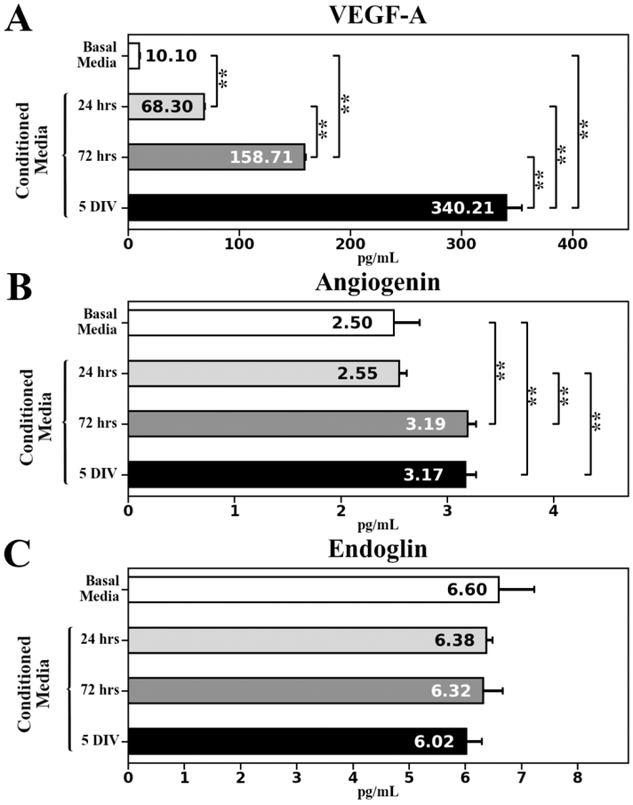
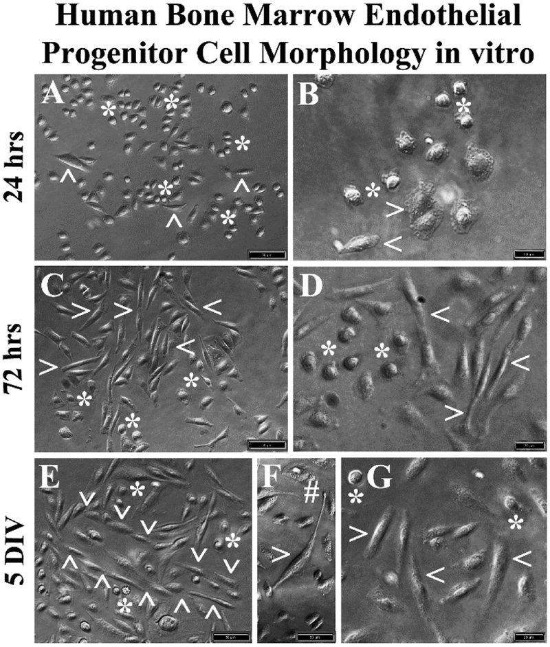
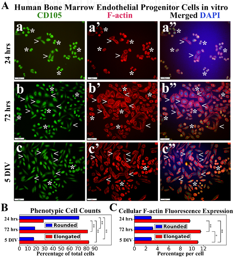
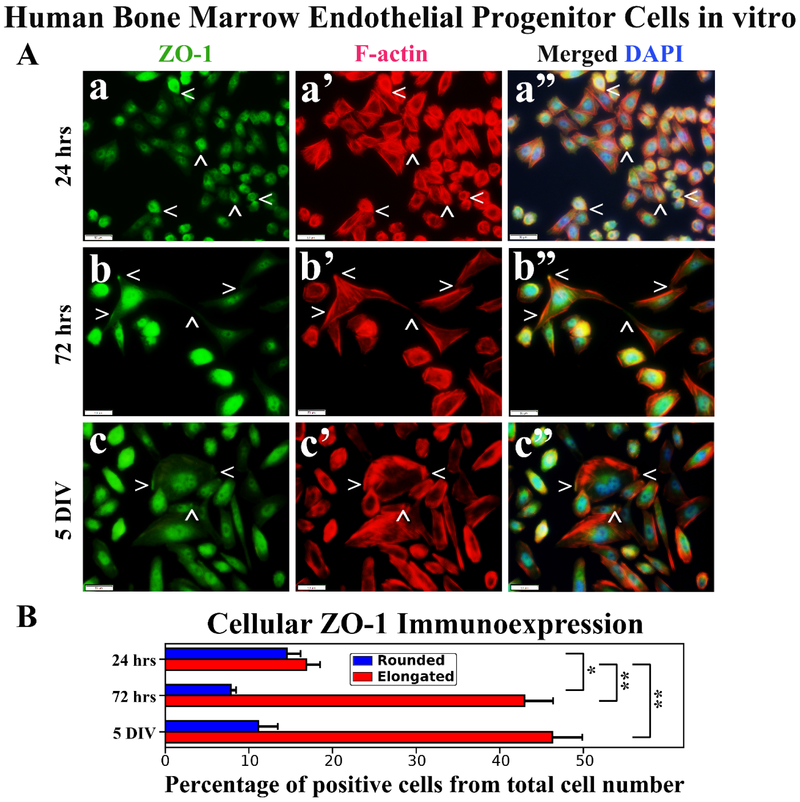
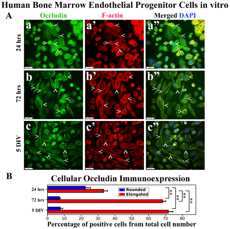
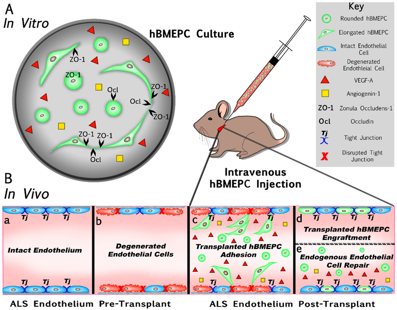
Amyotrophic lateral sclerosis (ALS) was recently recognized as a neurovascular disease. Accumulating evidence demonstrated blood-spinal-cord barrier (BSCB) impairment mainly via endothelial cell (EC) degeneration in ALS patients and animal models. BSCB repair may be a therapeutic approach for ALS. We showed benefits of human bone marrow endothelial progenitor cell (hBMEPC) transplantation into symptomatic ALS mice on barrier restoration; however, cellular mechanisms remain unclear. The study aimed to characterize hBMEPCs in vitro under normogenic conditions. hBMEPCs were cultured at different time points. Enzyme-linked immunosorbent assay (ELISA) was used to detect concentrations of angiogenic factors (VEGF-A, angiogenin-1, and endoglin) and angiogenic inhibitor endostatin in conditioned media. Double immunocytochemical staining for CD105, ZO-1, and occludin with F-actin was performed. Results showed predominantly gradual significant post-culture increases of VEGF-A and angiogenin-1 levels. Cultured cells displayed distinct rounded or elongated cellular morphologies and positively immunoexpressed for CD105, indicating EC phenotype. Cytoskeletal F-actin filaments were re-arranged according to cell morphologies. Immunopositive expressions for ZO-1 were detected near inner cell membrane and for occludin on cell membrane surface of adjacent hBMEPCs. Together, secretion of angiogenic factors by cultured cells provides evidence for a potential mechanism underlying endogenous EC repair in ALS through hBMEPC transplantation, leading to restored barrier integrity. Also, ZO-1 and occludin immunoexpressions, confirming hBMEPC interactions in vitro, may reflect post-transplant cell actions in vivo.

Keywords: Angiogenic factors; F-actin; Human bone marrow endothelial progenitor cells; In vitro; Tight junction proteins.


Conflict of interest statement

Conflict of Interest

The authors declare no conflict of interest.

Figures

Figure 1.
Figure 1.. Angiogenic factors ELISA analysis in conditioned media collected at different time points from cultured hBMEPCs.
(A) Significant gradual increase of VEGF-A level was detected in conditioned media from 24 hrs to 5 DIV of cultured hBMEPCs. (B) Significantly elevated angiogenin levels were determined in conditioned media at 72 hrs and 5 DIV of cultured hBMEPCs vs. 24 hrs or basal media. (C) There were no significant differences in endoglin levels between conditioned media at all measured time points and basal media. **p < 0.01.
Figure 2.
Figure 2.. Phase-contrast images of hBMEPCs in vitro at different time points.
(A, B) The hBMECs showed mainly morphologically rounded cells (asterisks) and a few elongated cells (arrowheads) were observed at 24 hrs of culture in basal media. (C, D) More elongated cells (arrowheads) were detected in cultures at 72 hrs. Numerous rounded cells (asterisks) were noticed. (E) At 5 DIV, a tubular-like vessel formation (arrowheads) composed of elongated cells was determined. (F) Elongated cells also displayed long processes with tip development (hashtag). (G) A few rounded cells (arrowheads) were still observed at 5 DIV. Scale bar in A, C, E, F is 50 μm; in B, D, G is 20 μm.
Figure 3.
Figure 3.. Immunocytochemical analyses of hBMEPCs in vitro at different time points for CD105 and F-actin expressions.
(A) CD105 immunoexpression was detected in all cells at each time point (green, a, b, c). During post-culture, F-actin immunoexpression (red) displayed diffusely within cytosol of rounded hBMEPCs (arrowheads) and elongated cells (arrowheads) exhibited organized filamentous strips in cell cytoplasm (a’, b’, c’). Merged images (a”, b”, c”) are shown with DAPI (blue). Scale bar in a-c” is 50 μm. (B) Rounded hBMEPC prevalence significantly decreased in conjunction with significantly increased occurrence of elongated hBMEPC from 24 hrs to 5 DIV. (C) Although F-actin immunoexpression levels remained stable over time, a significant increase of cytoskeletal filaments was determined in elongated cells from 24 hrs to 5 DIV. *p < 0.05, **p < 0.01.
Figure 4.
Figure 4.. Immunocytochemical analysis of hBMEPCs in vitro at different time points for ZO-1 and F-actin.
(A) Rounded hBMEPCs (a) displayed diffuse presence of ZO-1 (green, arrowheads) in contrast to ZO-1 localization in elongated hBMEPCs (b, c). ZO-1 immunoexpression was mainly detected within elongated cell filopodia tips (b) or near inner cell membrane (c). Merged images (a”, b”, c”) are shown with DAPI (blue). Scale bar in a-c” is 50 μm. (B) Rounded ZO-1 immunopositive hBMEPCs prevalence decreased significantly from 24 hrs to 72 hrs and no significant differences were detected between 72 hrs and 5 DIV cultures. Prevalence of ZO-1 immunopositive elongated hBMEPCs significantly increased across all time points. *p < 0.05, **p < 0.01.
Figure 5.
Figure 5.. Immunocytochemical analysis of hBMEPCs in vitro at different time points for occludin and F-actin.
(A) Occludin immunoexpression in rounded hBMEPCs localized on cell membrane surfaces and elongated cells showed this protein immunoexpression as strips between adjacent cells (green, arrowheads, a, b, c). Occludin immunoexpression appeared to be distal from F-actin in numerous cells (red, arrowheads, a’, b’, c’). Merged images (a”, b”, c”) are shown with DAPI (blue). Scale bar in a-c” is 50 μm. (B) Percentage of rounded occludin immunopositive hBMEPCs substantially decreased from 24 hrs to 5 DIV. Significantly increased percentage of elongated hBMEPCs immunoexpressing occludin was determined from 24 hrs to 5 DIV. **p < 0.01.
Figure 6.
Figure 6.. Schematic diagram summarizing hBMEPCs in vitro results and providing effective therapeutic mechanism(s) of transplanted cells into a mouse modeling ALS.
(A) hBMEPCs in vitro showed changes in morphological phenotype from rounded to elongated cells, secreted VEGF-A and angiogenin-1, and expressed tight junction ZO-1 and occludin proteins during post-culture. (B) Dysfunction of BBB/BSCB in ALS is mainly characterized by endothelial cells transition from an intact (a) to a degenerated (b) status. Intravenous transplantation of hBMEPCs into early symptomatic G93A SOD1 mice significantly repaired BSCB by widespread cell adhesion (c) followed by cell engraftment (d) into capillary lumen of the spinal cords and brains. The presence of administered cells in blood circulation of mice was also noted at four weeks after cell treatment. We hypothesized that hBMEPCs may not only contribute to endogenous restoration of damaged endothelium via secretion of angiogenic factors, but also facilitate proper maintenance of vascular homeostasis by increasing endothelial cell-cell “tightness” (e).

Similar articles

Cited by

References

    1. Adams RH, Alitalo K, 2007. Molecular regulation of angiogenesis and lymphangiogenesis. Nat. Rev. Mol. Cell Biol 8, 464–478. 10.1038/nrm2183 - DOI - PubMed
    1. Bataveljic D, Milosevic M, Radenovic L, Andjus P, 2014. Novel molecular biomarkers at the blood-brain barrier in ALS. Biomed. Res. Int 2014, 907545 10.1155/2014/907545 - DOI - PMC - PubMed
    1. Bataveljić D, Nikolić L, Milosević M, Todorović N, Andjus PR, 2012. Changes in the astrocytic aquaporin-4 and inwardly rectifying potassium channel expression in the brain of the amyotrophic lateral sclerosis SOD1(G93A) rat model. Glia 60, 1991–2003. 10.1002/glia.22414 - DOI - PubMed
    1. Blancas AA, Wong LE, Glaser DE, McCloskey KE, 2013. Specialized tip/stalk-like and phalanx-like endothelial cells from embryonic stem cells. Stem Cells Dev. 22, 1398–1407. 10.1089/scd.2012.0376 - DOI - PMC - PubMed
    1. Boillée S, Vande Velde C, Cleveland DW, 2006. ALS: a disease of motor neurons and their nonneuronal neighbors. Neuron 52, 39–59. 10.1016/j.neuron.2006.09.018 - DOI - PubMed

Publication types