Review
. 2020 Mar:75:100777.
doi: 10.1016/j.preteyeres.2019.100777. Epub 2019 Sep 4.

Membrane-associated mucins of the ocular surface: New genes, new protein functions and new biological roles in human and mouse

Affiliations
Review

Membrane-associated mucins of the ocular surface: New genes, new protein functions and new biological roles in human and mouse

M Elizabeth Fini et al. Prog Retin Eye Res. 2020 Mar.

Abstract

The mucosal glycocalyx of the ocular surface constitutes the point of interaction between the tear film and the apical epithelial cells. Membrane-associated mucins (MAMs) are the defining molecules of the glycocalyx in all mucosal epithelia. Long recognized for their biophysical properties of hydration, lubrication, anti-adhesion and repulsion, MAMs maintain the wet ocular surface, lubricate the blink, stabilize the tear film and create a physical barrier to the outside world. However, it is increasingly appreciated that MAMs also function as cell surface receptors that transduce information from the outside to the inside of the cell. A number of excellent review articles have provided perspective on the field as it has progressed since 1987, when molecular cloning of the first MAM was reported. The current article provides an update for the ocular surface, placing it into the broad context of findings made in other organ systems, and including new genes, new protein functions and new biological roles. We discuss the epithelial tissue-equivalent with mucosal differentiation, the key model system making these advances possible. In addition, we make the first systematic comparison of MAMs in human and mouse, establishing the basis for using knockout mice for investigations with the complexity of an in vivo system. Lastly, we discuss findings from human genetics/genomics, which are providing clues to new MAM roles previously unimagined. Taken together, this information allows us to generate hypotheses for the next stage of investigation to expand our knowledge of MAM function in intracellular signaling and roles unique to the ocular surface.

Keywords: Epithelial tissue-equivalent; Glycocalyx; Knockout mouse; Membrane-associated mucin; Ocular surface; Signal transduction.


Conflict of interest statement

Declaration of Interest

MEF and SJ are named as inventors on an issued United States patent entitled “Structure/Function of Clusterin Pharmaceuticals” and a pending patent application entitled “Method to Protect and Seal the Ocular Surface” (United States application 16/103,741, filed Aug 14, 2018), assigned to the University of Southern California and related to work mentioned herein. MEF is a co-founder and serves as Chief Scientific Officer for Proteris Biotech, Inc., a company focused on developing pharmaceuticals for treating eye disease. The other authors have no commercial or proprietary interest in any concept or product described in this article.

Figures

Figure 1.
Figure 1.. Mucin gene expression in human conjunctival epithelium.
Microarray analysis of impression cytology samples indicates that MUC20 is the most highly expressed mucin gene in human conjunctiva. n.d.: not detected. CD164 was previously designated as MUC24. MUC21 and MUC22 are not included in this analysis. From (Woodward and Argueso, 2014), with permission.
Figure 2.
Figure 2.. Location of Genes for MUC 21 and MUC22 at Chromosomal Region 6p21.32–33 and Expression in the Corneal Epithelium.
Top: Schematic of chromosomal region 6p21.32–33 from NCBI Gene depicting annotated genes surrounding an identified quantitative trait locus (QTL) for steroid-induced ocular hypertension (red arrow) in the transcriptional promotor region of HCG22. Bottom: Total RNA was purified from cultured primary human corneal epithelial cells (HCE) and cells of the trabecular meshwork (TBM) cell line TM-1, and used for cDNA synthesis. RT-PCR using the cDNA was performed using specific primers from MUC21, MUC22, and HCG22; the products were resolved on a 1.5% agarose gel. Primers were designed to detect only the coding transcript. Similar results were obtained using three primary TBM cell lines (not shown). RTase: reverse transcriptase; HCE: primary corneal epithelial cells obtained from corneal rims. From (Jeong et al., 2015) with permission.
Figure 3.
Figure 3.. Immunolocalization of MUC21 and MUC22 in the Human Corneal Epithelium and the Human Lacrimal Gland.
An anterior segment isolated from a human donor eye was formalin-fixed within 24-hours post-mortem and paraffin-embedded. A formalin-fixed human lacrimal gland embedded in paraffin was obtained from the Ophthalmic Pathology Laboratory of Tufts Medical Center. Tissues cross-sections were prepared, then processed and indirectly immunostained for MUC21 or MUC22 as described (Itakura et al., 2019). The human MUC21 primary antibody was purchased from Sigma-Aldrich Corp. (St. Louis, MO). It is derived from a rabbit polyclonal antisera raised against a peptide from the human MUC21 cytoplasmic tail (561-CVRNSLSLRN TFNTAVYHPH GLNHGLGPGP GGNHGAPHRP RWSPNWFWRR PVSSIAMEMS GRNS-624), then affinity-purified. The human MUC22 primary antibody was characterized in one of our labs, as described (Hijikata et al., 2011). A rabbit polyclonal antisera produced by GENENET (Fukuoka, Japan) was raised against a peptide (TPTNVIKPSGYLQP) from the human MUC22 stem region located just before the transmembrane domain, then affinity-purified. A 3,3′-diaminobenzidine (DAB) chromogen kit was used to detect secondary antibody binding (Vector Laboratories, Burlingame, CA). The negative control (Neg. control) omitted the primary antibody. Sections were counterstained with hematoxylin. A-C) Cross-sections through the anterior segment focusing on immunostaining results (brown color) in the cornea epithelium. The hematoxylin counterstain is dark blue. Magnification = 40X. D-L) Cross-sections through the lacrimal showing immunostaining results (brown color). The hematoxylin counterstain is dark blue. D-F) Low magnification view (10X); G-I) Higher magnification (40X) focusing on a lacrimal duct; J-L) Higher magnification focusing on serous acini. These experimental findings have not been previously published.
Figure 4.
Figure 4.. Prototype of a Membrane Associated Mucin (MAM).
The graphic depicts a prototypical MAM, the structure of which is similar to a classic, single-pass transmembrane immune receptor. A signal peptide motif is found at the N-terminal of the precursor polypeptide chain to enable its membrane insertion; it may be retained in the mature protein (1). The mature protein is composed of two subunits that self-associate, arising from intracellular cleavage. The large subunit is entirely extracellular and contains the VNTR. The small subunit consists of a short extracellular region, a single-pass transmembrane domain, and a cytoplasmic tail (CT). The large subunit of the MAM, together with the extracellular portion of the small subunit, comprise the extracellular domain (ED). The ED also contains conserved sequence motifs as modular elements such as the Sperm protein, Enterokinase and Agrin module (SEA) and EGF-like modules.
Figure 5.
Figure 5.. Proposed Evolutionary Subgroupings of Epithelial Membrane Associated Mucins (MAMs).
The best evidence is that MAMs arose largely through a process of convergent evolution, but they can be grouped into evolutionarily-related subgroups based on their genetic backgrounds, as shown in the graphic. The rationale for the groupings is discussed in the text. Some of the information in this graphic is summarized from (Duraisamy et al., 2006). The complete analysis shown here has not been previously published.
Figure 6.
Figure 6.. Modular Architecture of Ocular Surface Membrane Associated Mucins (MAMs).
Shown are the extended conformations of MAM proteins prior to intracellular processing, but with the final relationship to the plasma membrane depicted. The extracellular domain of each protein is to the left of the plasma membrane. MAMs could not be drawn to scale because of extreme size differences, but an effort was made to depict relative differences in overall size, and relative location and sizes of the modular units. The signal peptides are located at the amino-terminus of each protein. The approximate intracellular cleavage sites of each mucin are indicated by scissors. MUC20 has been experimentally determined to associate with the plasma membrane, but no transmembrane domain has been identified. SP: signal peptide; TM: transmembrane domain; VNTR: Variable Number Tandem Repeats; conserved modular domains as in Table 3. GSLV: proposed cleavage site for MUC21.
Figure 7.
Figure 7.. Sequence Analysis of Membrane-Associated Mucin (MAM) Cytoplasmic Tails (CTs).
Amino acid sequences of the human MAM CTs, as determined by conceptual translation of the mRNA sequence, are shown. At the end of each sequence, the amino acid count is indicated. If there is an orthologue in mouse and rat, this is also shown and conserved amino acids are identified with a line between the two sequences. Lower case letters in red indicate serine, threonine or tyrosine residues confirmed experimentally to be phosphorylated on the PhosphoSitePlus website. Lower case letters in blue indicate serine, threonine or tyrosine residues predicted by the NetPhos 3.1 Server. Some of the many confirmed MUC1 interacting proteins are indicated in red above the recognition sequence: serine-threonine kinase GSK3B (SXXXS); tyrosine kinase PIK3R1 (regulatory subunit; Y20HPM); receptor tyrosine kinase EGFR (Y46EKV/Y46EEV); phospholipase PLCG1 (Y35VPP); adherens junction component beta-catenin CTNNB1 (SXXXXXSSL); adaptor protein GRB2 (Y60TNP); tyrosine kinase SRC (Y46EK/EV). Predicted phosphorylating kinase are indicated in blue above the predicted phosphorylation site. If there are additional predictions for the mouse/rat sequences, these are indicated as well. Predicted phosphorylating kinases: gsk: glycogen synthase kinase-3 isoform; pka: protein kinase A isoforms; pkc: protein kinase C isoforms. Others designated by HUGO nomenclature. The proposed N-terminal pamitoylation site in MUC1 and adjacent polybasic amino acid stretches in MUC1 and MUC16 are in blue text and underlined. Regions predicted to have disordered protein binding properties in human MUC21 and MUC22 are in underlined black text. This compilation, with its new analyses, has not been previously published.
Fig 8.
Fig 8.. Application of Oxidative Stress to HCLE Epithelial-Equivalents with Mucosal Differentiation.
Islands of cells with mucosal differentiation at the surface of HCLE epithelial-equivalents exclude rose bengal (A and B). When oxidative stress is applied (10 mM tBHP in DMEM/F12 medium for 2 hours, as previously described (Webster et al., 2018)), many of these cells lose their transcellular barrier function and rose bengal penetrates (C and D). Rose Bengal staining was performed by incubating the cells 5 minutes in 0.1% Rose Bengal as previously described (Argueso et al., 2006). Magnification: A and C are 4x; B and D are 10x. This example has not been previously published, but is similar to findings published in (Webster et al., 2018)
Figure 9.
Figure 9.. Schematic of the Mouse Ocular Surface System.
A) Eye in the mouse showing positioning of the glands; B) Larger and side view of A; C) Larger view of the isolated eye cross section. ELG: Extraorbital Lacrimal Gland; ILG: Intraorbital Lacrimal Gland; HG: Harderian Gland
Figure 10.
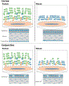
Figure 10.. Comparison of MUC1, MUC4, and MUC16 Localization at the Human and Mouse Ocular Surface.
The graphics compare the corneal epithelium (top) and conjunctiva (bottom) from human (left) and mouse (right). As depicted, the corneal epithelium in mouse has more cell layers than human; the human corneal stroma is thicker than that of mouse. The expanded insets depict a single apical epithelia cell from the corneal or conjunctival epithelium of mouse or human, showing the surface microplicae. With the EDs of MAMs MUC1, MUC4, and MUC16 projecting outward into the tear film. The EDs of the two longest MAMs, MUC16 and MUC4, are substantially shorter in mouse than human. MUC4 appears to substitute for MUC16 on the corneal epithelium of mouse, which further reduces the overall length of MAM EDs on the corneal epithelium. MUC1: orange; MUC4: blue; MUC16: green.

Similar articles

Cited by

References

    1. Abelson MB, Ingerman A, 2005. The Dye-namics of Dry-Eye Diagnosis. Review of Ophthalmology https://www.reviewofophthalmology.com/article/the-dye-namics-of-dry-eye-....
    1. Ablamowicz AF, Nichols JJ, 2016. Ocular Surface Membrane-Associated Mucins. The ocular surface 14, 331–341. - PubMed
    1. Agrawal B, Krantz MJ, Parker J, Longenecker BM, 1998. Expression of MUC1 mucin on activated human T cells: implications for a role of MUC1 in normal immune regulation. Cancer research 58, 4079–4081. - PubMed
    1. Aithal A, Junker WM, Kshirsagar P, Das S, Kaur S, Orzechowski C, Gautam SK, Jahan R, Sheinin YM, Lakshmanan I, Ponnusamy MP, Batra SK, Jain M, 2018. Development and characterization of carboxy-terminus specific monoclonal antibodies for understanding MUC16 cleavage in human ovarian cancer. PloS one 13, e0193907. - PMC - PubMed
    1. Al-Bataineh MM, Sutton TA, Hughey RP, 2017. Novel roles for mucin 1 in the kidney. Curr Opin Nephrol Hypertens 26, 384–391. - PMC - PubMed

Publication types