. 2020 Jan:23:102090.
doi: 10.1016/j.nano.2019.102090. Epub 2019 Sep 4.

Bioactive proteins delivery through core-shell nanofibers for meniscal tissue regeneration

Affiliations

Bioactive proteins delivery through core-shell nanofibers for meniscal tissue regeneration

Jihye Baek et al. Nanomedicine. 2020 Jan.

Abstract

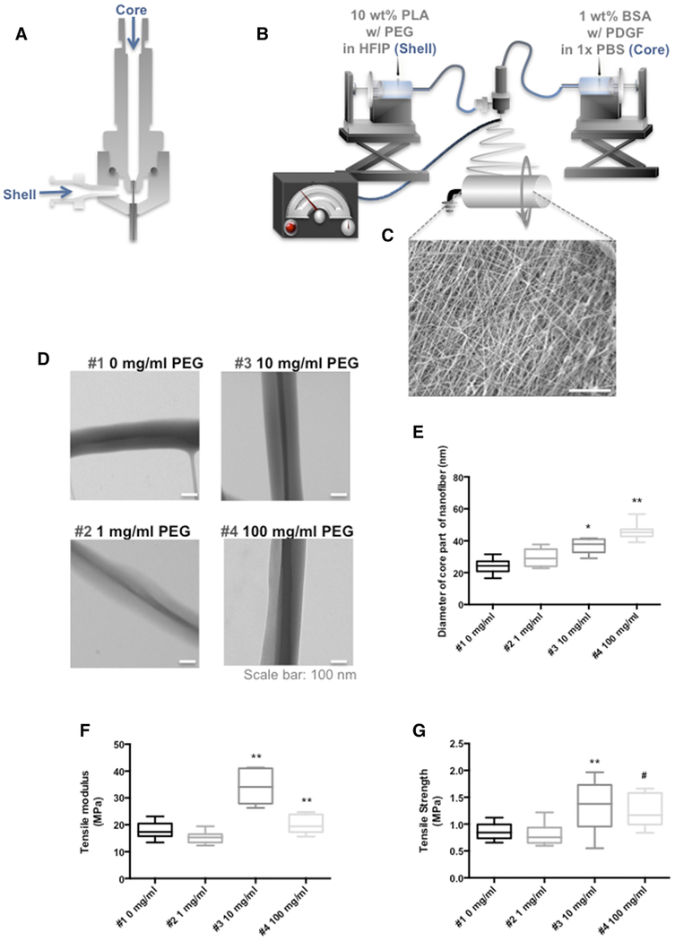
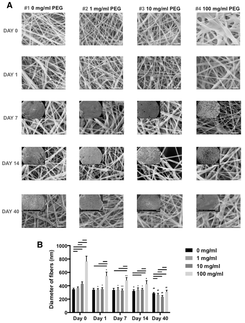
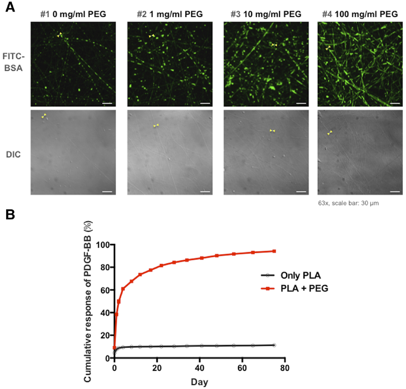
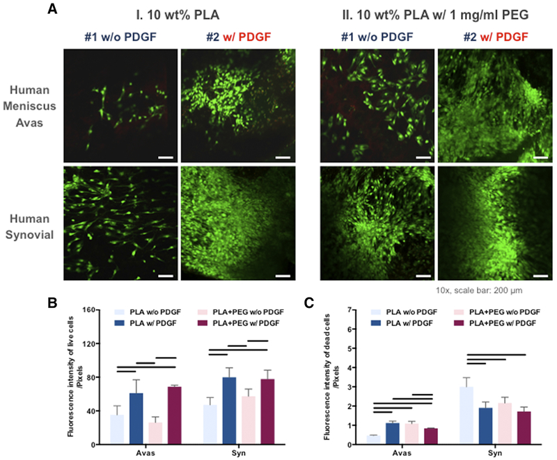
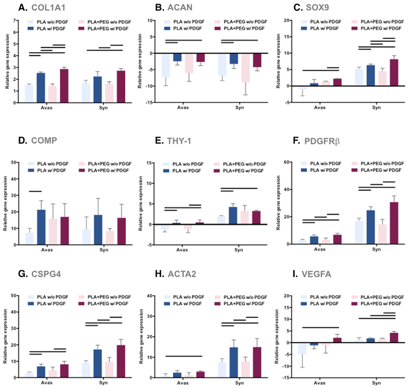
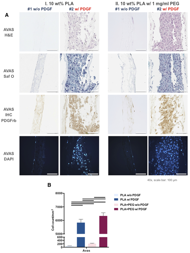
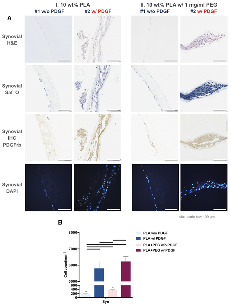
Mimicking the ultrastructural morphology of the meniscus with nanofiber scaffolds, coupled with controlled growth-factor delivery to the appropriate cells, can help engineer tissue with the potential to grow, mature, and regenerate after in vivo implantation. We electrospun nanofibers encapsulating platelet-derived growth factor (PDGF-BB), which is a potent mitogen and chemoattractant in a core of serum albumin contained within a shell of polylactic acid. We controlled the local PDGF-BB release by adding water-soluble polyethylene glycol to the polylactic acid shell to serve as a porogen. The novel core-shell nanofibers generated 3D scaffolds with an interconnected macroporous structure, with appropriate mechanical properties and with high cell compatibility. Incorporating PDGF-BB increased cell viability, proliferation, and infiltration, and upregulated key genes involved in meniscal extracellular matrix synthesis in human meniscal and synovial cells. Our results support proof of concept that these core-shell nanofibers can create a cell-favorable nanoenvironment and can serve as a system for sustained release of bioactive factors.

Keywords: Co-axial electrospinning; Core-shell structure; Meniscus; Nanofibers; PDGF-BB; Tissue engineering.


Conflict of interest statement

Disclosures: The authors have no conflicts of interest to declare.

Figures

Figure 1.
Figure 1.. Co-axial electrospinning and structural properties of co-axial electrospun nanofibers.
(A) Schematic illustration of a co-axial electrospinning spinneret used to prepare core-shell nanofibers. (B) Overview of the core-shell electrospinning equipment. (C) SEM image of co-axial electrospun scaffold (Mag: 1250x; scale bar 20 μm). (D) Transmission electron microscopy images illustrating the presence of BSA cores in PLA shells with 0, 1, 10, and 100 mg/ml PEG (Mag: 30,000x; scale bar: 100 nm) (E) Average diameter of the BSA core increased with increasing concentrations of PEG. *= P < 0.05 compared to 0, and 1 mg/ml, **= P < 0.05 compared to 0, 1, 10 mg/ml (n = 10 per condition) (F) Tensile modulus and (G) ultimate tensile strength of core-shell nano fibrous scaffolds with different concentration of PEG (0, 1, 10, and 100 mg/ml). **= P < 0.05 compared to 0, and 1 mg/ml, #= P < 0.05 compared to 10 mg/ml. (n = 10 per condition)
Figure 2.
Figure 2.. Size distribution of core-shell electrospun fibers with different PLA/PEG formulations over 40 days in phosphate buffered solution.
(A) SEM images of fibers (10% PLA + 0, 1, 10, or 100 mg/ml PEG) at day 0, 1, 7, 14, and 40 (Mag: 10,000x; scale bar: 5 μm). Insert: SEM images of each specimen (10% PLA + 0, 1, 10, or 100 mg/ml PEG) at day 7, 14, and 40 (Mag: 35x; scale bar: 500 μm) (B) Diameters of electrospun PLA nanofibers with different concentration of PEG cultivated in PBS over 40 days (electrospun fibers were examined by SEM and fiber size measured using ImageJ). Line= P < 0.05 between groups, *= P < 0.05 compared to 0 day, **= P < 0.05 compared to 0, and 1 day, #= P < 0.05 compared to 0, 1, and 7 day, ##= P < 0.05 compared to 0, 1, 7, and 14 day, += P < 0.05 compared to 0, and 7 day.
Figure 3.
Figure 3.. Encapsulation of FITC-BSA and PDGF-BB release from electrospun nanofibers.
(A) Laser confocal microscope and DIC images of core-shell nanofibers with FITC-BSA in formulations of PEG-blended PLA. (B) Controlled release of encapsulated PDGF-BB from PLA and PLA-PEG core-shell scaffolds. (10% PLA + 1 mg/ml PEG).
Figure 4.
Figure 4.. Cellular response of the human meniscus avascular and synovial cells cultured on PLA nanofibers with or without PDGF and with or without PEG.
(A) Confocal microscope images of human meniscus and synovial cells cultured on aligned PLA nanofibers demonstrating viability in confocal images (Mag. 10x; scale bar: 200 mm). (B) Quantitative analysis of fluorescent intensity of the live and dead meniscus and synovial cells in core-shell nanofibrous scaffolds (n = 3 donors, 3 replicates). Line= P < 0.05 between groups.
Figure 5.
Figure 5.. Gene expression levels of human meniscus avascular cells and synovial cells cultured on PLA scaffolds were compared to cells in monolayer culture.
Shown here are differential effects of PDGF-BB and PEG (3 donors, 3 replicates). (A) COL1A1 gene expression, (B) ACAN gene expression, (C) SOX9 gene expression, (D) COMP gene expression, (E) THY-1 gene expression, (F) PDGFRβ gene expression, (G) CSPG4 gene expression, (H) ACTA2 gene expression, and (I) VEGFA gene expression relative to monolayer controls. Line= P < 0.05 between groups.
Figure 6.
Figure 6.. PDGF-BB significantly increased infiltration of human meniscus avascular cells into the scaffolds.
(A) Representative histologic images of core-shell nanofibers with human meniscus avascular cells: H&E, Safranin O Fast Green, DAPI, and PDGFRβ immunostaining (Mag.: 40x; scale bar: 100 μm). (B) Number of meniscus avas cells infiltrated into core-shell nanofibers (3 donors, 3 replicates). Line= P < 0.05 between groups.
Figure 7.
Figure 7.. PDGF-BB significantly increased infiltration of human synovial cells into the scaffolds.
(A) Representative histologic images of core-shell nanofibers with human synovial cells: H&E, Safranin O Fast Green, DAPI, and PDGFRβ immunostaining (Mag.: 40x; scale bar: 100 μm). (B) Number of synovial cells infiltrated into core-shell nanofibers. Line= P < 0.05 between groups, *=P < 0.05 between each cell type.

Similar articles

Cited by

References

    1. Benjamin M, Ralphs JR. Fibrocartilage in tendons and ligaments — an adaptation to compressive load. J Anat 1998;193:481–94. - PMC - PubMed
    1. Petersen W, Tillmann B. Collagenous fibril texture of the human knee joint menisci. Anat Embryol (Berl) 1998;197:317–24. 10.1007/s004290050141 - DOI - PubMed
    1. Hickey DS, Hukins DW. Relation between the structure of the annulus fibrosus and the function and failure of the intervertebral disc. Spine (Phila Pa 1976) 1980;5:106–16. - PubMed
    1. Lee KI, Olmer M, Baek J, D’Lima DD, Lotz MK. Platelet-derived growth factor-coated decellularized meniscus scaffold for integrative healing of meniscus tears. Acta Biomater 2018;76:126–34. 10.1016/j.actbio.2018.06.021 - DOI - PMC - PubMed
    1. Baek J, Sovani S, Choi W, Jin S, Grogan SP, D’Lima DD. Meniscal Tissue Engineering Using Aligned Collagen Fibrous Scaffolds: Comparison of Different Human Cell Sources. Tissue Eng Part A 2018;24:81–93. 10.1089/ten.TEA.2016.0205 - DOI - PMC - PubMed

Publication types