

Structural characterization and biological function of bivalent binding of CD2AP to intrinsically disordered domain of chikungunya virus nsP3 protein
- PMID: 31493651
- PMCID: PMC7144582
- DOI: 10.1016/j.virol.2019.08.022
Structural characterization and biological function of bivalent binding of CD2AP to intrinsically disordered domain of chikungunya virus nsP3 protein
Abstract
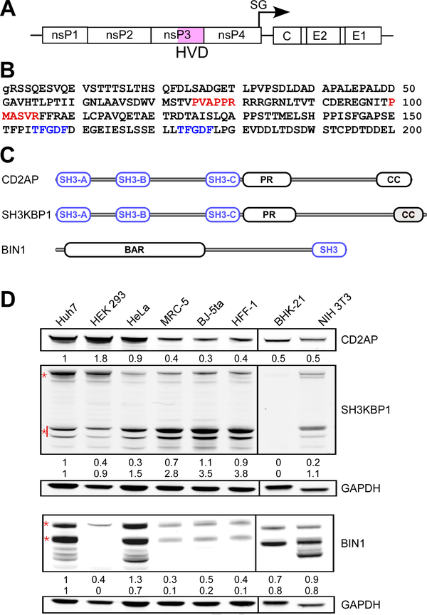
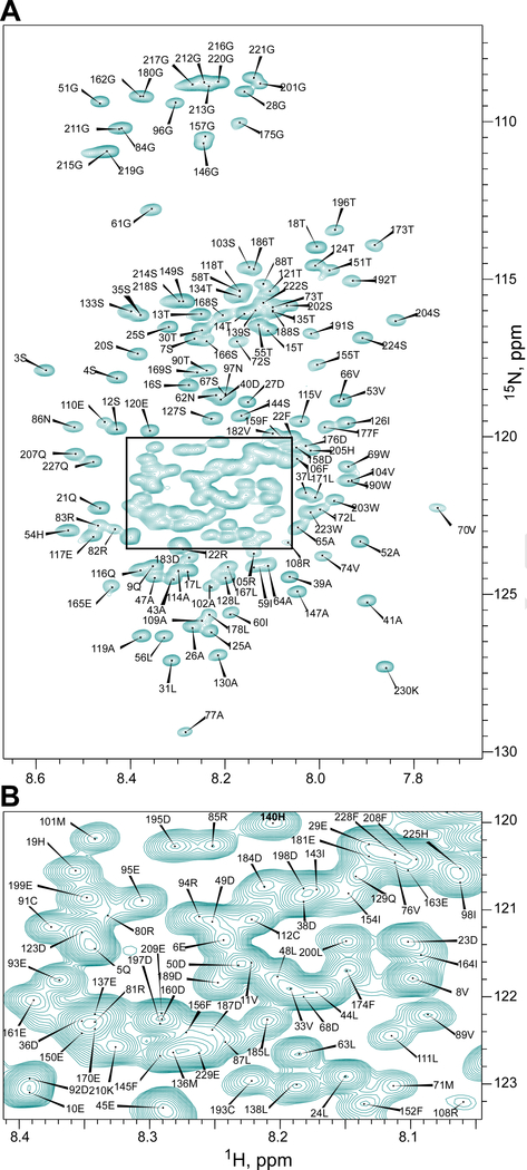
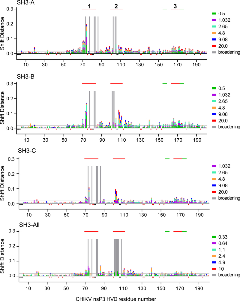
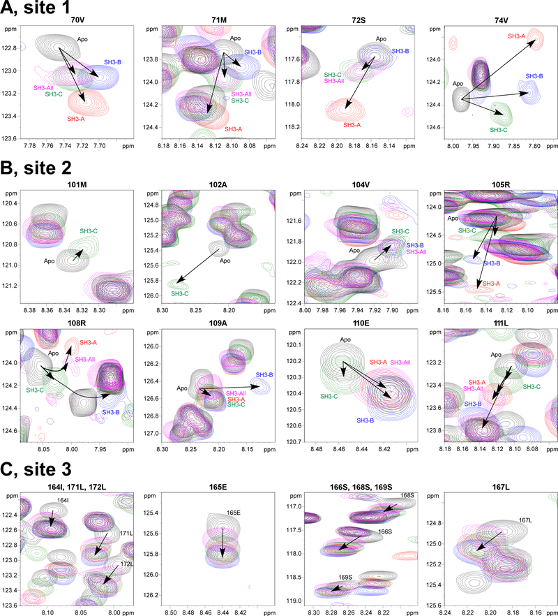
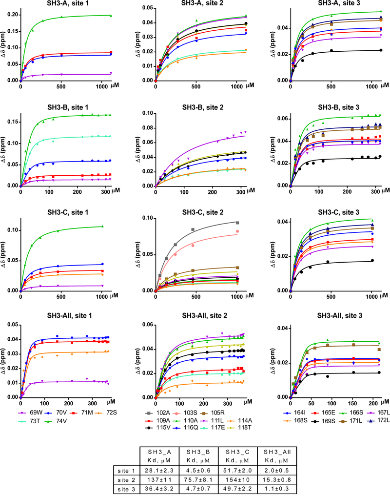
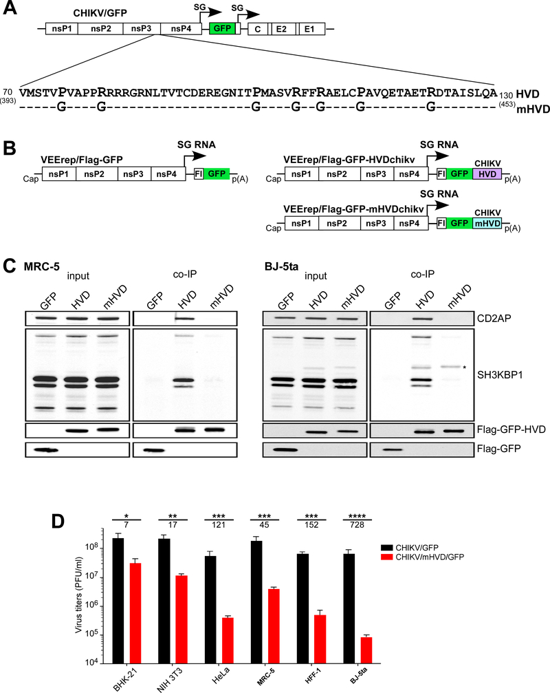
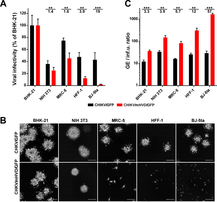
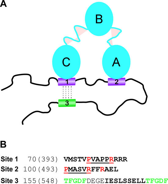
Alphavirus nsP3 proteins contain long, intrinsically disordered, hypervariable domains, HVD, which serve as hubs for interaction with many cellular proteins. Here, we have deciphered the mechanism and function of HVD interaction with host factors in alphavirus replication. Using NMR spectroscopy, we show that CHIKV HVD contains two SH3 domain-binding sites. Using an innovative chemical shift perturbation signature approach, we demonstrate that CD2AP interaction with HVD is mediated by its SH3-A and SH3-C domains, and this leaves the SH3-B domain available for interaction with other cellular factor(s). This cooperative interaction with two SH3 domains increases binding affinity to CD2AP and possibly induces long-range allosteric effects in HVD. Our data demonstrate that BIN1, CD2AP and SH3KBP1 play redundant roles in initiation of CHIKV replication. Point mutations in both CHIKV HVD binding sites abolish its interaction with all three proteins, CD2AP, BIN1 and SH3KBP1. This results in strong inhibition of viral replication initiation.
Keywords: Alphaviruses; BIN1; CD2AP; Chikungunya virus; Intrinsically disordered proteins; NMR; SH3 domain-containing proteins; SH3KBP1; Viral replication; nsP3.
Copyright © 2019 The Authors. Published by Elsevier Inc. All rights reserved.
Conflict of interest statement
Conflict of Interest
The authors have no conflict of interest to declare.
Figures
Similar articles
-
The Putative Roles and Functions of Indel, Repetition and Duplication Events in Alphavirus Non-Structural Protein 3 Hypervariable Domain (nsP3 HVD) in Evolution, Viability and Re-Emergence.Viruses. 2021 May 28;13(6):1021. doi: 10.3390/v13061021. Viruses. 2021. PMID: 34071712 Free PMC article. Review.
-
Structural and Functional Characterization of Host FHL1 Protein Interaction with Hypervariable Domain of Chikungunya Virus nsP3 Protein.J Virol. 2020 Dec 9;95(1):e01672-20. doi: 10.1128/JVI.01672-20. Print 2020 Dec 9. J Virol. 2020. PMID: 33055253 Free PMC article.
-
Mutation of CD2AP and SH3KBP1 Binding Motif in Alphavirus nsP3 Hypervariable Domain Results in Attenuated Virus.Viruses. 2018 Apr 27;10(5):226. doi: 10.3390/v10050226. Viruses. 2018. PMID: 29702546 Free PMC article.
-
Multiple Host Factors Interact with the Hypervariable Domain of Chikungunya Virus nsP3 and Determine Viral Replication in Cell-Specific Mode.J Virol. 2018 Jul 31;92(16):e00838-18. doi: 10.1128/JVI.00838-18. Print 2018 Aug 15. J Virol. 2018. PMID: 29899097 Free PMC article.
-
The Enigmatic Alphavirus Non-Structural Protein 3 (nsP3) Revealing Its Secrets at Last.Viruses. 2018 Feb 28;10(3):105. doi: 10.3390/v10030105. Viruses. 2018. PMID: 29495654 Free PMC article. Review.
Cited by
-
Identification of a non-canonical G3BP-binding sequence in a Mayaro virus nsP3 hypervariable domain.Front Cell Infect Microbiol. 2022 Aug 11;12:958176. doi: 10.3389/fcimb.2022.958176. eCollection 2022. Front Cell Infect Microbiol. 2022. PMID: 36034716 Free PMC article.
-
Venezuelan Equine Encephalitis Virus nsP3 Phosphorylation Can Be Mediated by IKKβ Kinase Activity and Abrogation of Phosphorylation Inhibits Negative-Strand Synthesis.Viruses. 2020 Sep 13;12(9):1021. doi: 10.3390/v12091021. Viruses. 2020. PMID: 32933112 Free PMC article.
-
The Putative Roles and Functions of Indel, Repetition and Duplication Events in Alphavirus Non-Structural Protein 3 Hypervariable Domain (nsP3 HVD) in Evolution, Viability and Re-Emergence.Viruses. 2021 May 28;13(6):1021. doi: 10.3390/v13061021. Viruses. 2021. PMID: 34071712 Free PMC article. Review.
-
Assigned NMR backbone resonances of the ligand-binding region domain of the pneumococcal serine-rich repeat protein (PsrP-BR) reveal a rigid monomer in solution.Biomol NMR Assign. 2020 Oct;14(2):195-200. doi: 10.1007/s12104-020-09944-9. Epub 2020 Apr 20. Biomol NMR Assign. 2020. PMID: 32314099 Free PMC article.
-
Overview on Chikungunya Virus Infection: From Epidemiology to State-of-the-Art Experimental Models.Front Microbiol. 2021 Oct 5;12:744164. doi: 10.3389/fmicb.2021.744164. eCollection 2021. Front Microbiol. 2021. PMID: 34675908 Free PMC article. Review.
References
-
- Ababou A, Pfuhl M, Ladbury JE, 2009. Novel insights into the mechanisms of CIN85 SH3 domains binding to Cbl proteins: solution-based investigations and in vivo implications. J Mol Biol 387, 1120–1136. - PubMed
-
- Dikic I, 2002. CIN85/CMS family of adaptor molecules. FEBS Lett 529, 110–115. - PubMed
-
- Favier A, Brutscher B, 2011. Recovering lost magnetization: polarization enhancement in biomolecular NMR. J Biomol NMR 49, 9–15. - PubMed
-
- Feuerstein S, Solyom Z, Aladag A, Favier A, Schwarten M, Hoffmann S, Willbold D, Brutscher B, 2012. Transient structure and SH3 interaction sites in an intrinsically disordered fragment of the hepatitis C virus protein NS5A. J Mol Biol 420, 310–323. - PubMed
Publication types
MeSH terms
Substances
Grants and funding
LinkOut - more resources
Full Text Sources
Medical
Molecular Biology Databases
Miscellaneous
