. 2019 Sep 7;12(1):53.
doi: 10.1186/s13072-019-0300-y.

TH2BS11ph histone mark is enriched in the unsynapsed axes of the XY body and predominantly associates with H3K4me3-containing genomic regions in mammalian spermatocytes

Affiliations

TH2BS11ph histone mark is enriched in the unsynapsed axes of the XY body and predominantly associates with H3K4me3-containing genomic regions in mammalian spermatocytes

Iyer Aditya Mahadevan et al. Epigenetics Chromatin. .

Abstract

Background: TH2B is a major histone variant that replaces about 80-85% of somatic H2B in mammalian spermatocytes and spermatids. The post-translational modifications (PTMs) on TH2B have been well characterised in spermatocytes and spermatids. However, the biological function(s) of these PTMs on TH2B have not been deciphered in great detail. In our attempt to decipher the unique function(s) of histone variant TH2B, we detected the modification in the N-terminal tail, Serine 11 phosphorylation on TH2B (TH2BS11ph) in spermatocytes.

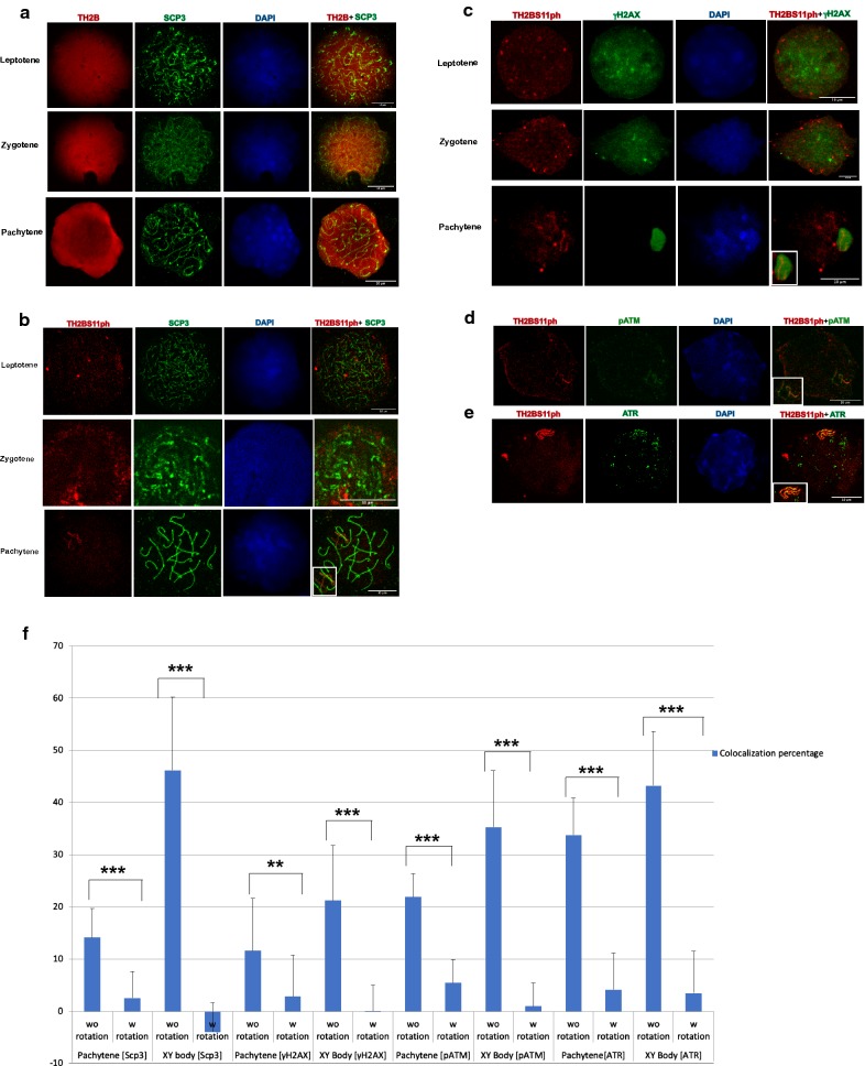
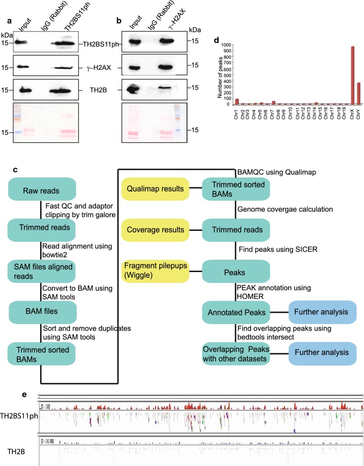
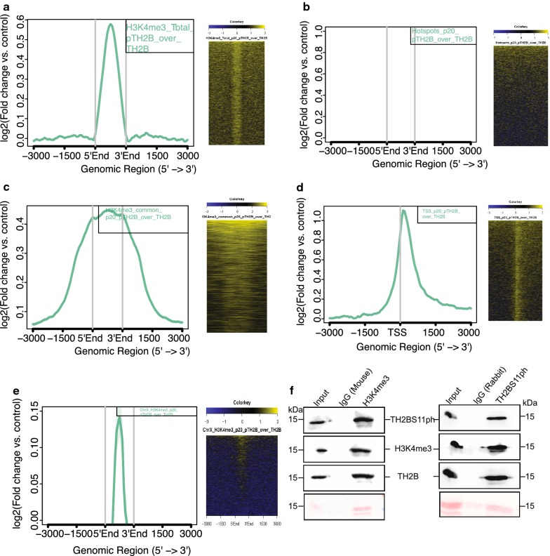
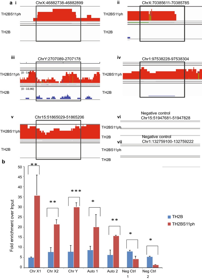
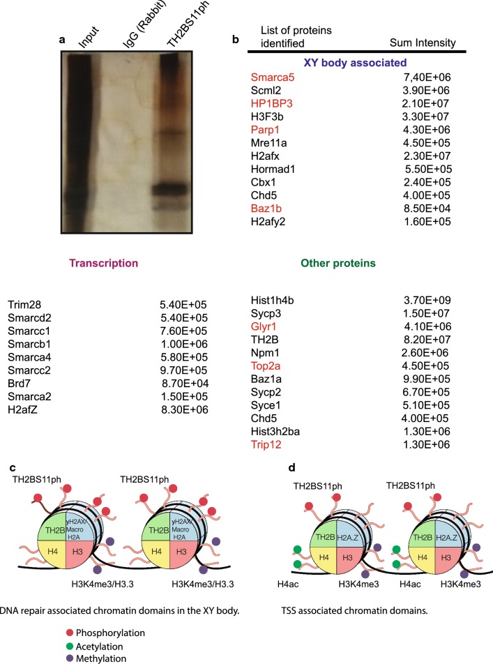
Results: The current study is aimed at understanding the function of the TH2BS11ph modification in the context of processes that occur during meiotic prophase I. Immunofluorescence studies with the highly specific antibodies revealed that TH2BS11ph histone mark is enriched in the unsynapsed axes of the sex body and is associated with XY body-associated proteins like Scp3, γH2AX, pATM, ATR, etc. Genome-wide occupancy studies as determined by ChIP sequencing experiments in P20 C57BL6 mouse testicular cells revealed that TH2BS11ph is enriched in X and Y chromosomes confirming the immunofluorescence staining pattern in the pachytene spermatocytes. Apart from the localisation of this modification in the XY body, TH2BS11ph is majorly associated with H3K4me3-containing genomic regions like gene promoters, etc. These data were also found to corroborate with the ChIP sequencing data of TH2BS11ph histone mark carried out in P12 C57BL6 mouse testicular cells, wherein we found the predominant localisation of this modification at H3K4me3-containing genomic regions. Mass spectrometry analysis of proteins that associate with TH2BS11ph-containing mononucleosomes revealed key proteins linked with the functions of XY body, pericentric heterochromatin and transcription.

Conclusions: TH2BS11ph modification is densely localised in the unsynapsed axes of the XY body of the pachytene spermatocyte. By ChIP sequencing studies in mouse P12 and P20 testicular cells, we demonstrate that TH2BS11ph is predominantly associated with H3K4me3 positive genomic regions like gene promoters, etc. We propose that TH2BS11ph modification could act alone or in concert with other histone modifications to recruit the appropriate transcription or XY body recombination protein machinery at specific genomic loci.

Keywords: Coimmunoprecipitation; H3K4me3 co-association; Immunofluorescence; Spermatocytes; TH2B; TH2B serine 11 phosphorylation.


Conflict of interest statement

The authors declare that they have no competing interests.

Figures

Fig. 1
Fig. 1
Identification of TH2BS11ph modification by mass spectrometry and characterization of the TH2BS11ph antibody. a Genome-wide replacement of H2B by TH2B histone variant in mammalian spermatocytes and spermatids [12]. b Model of the TH2B-containing nucleosome highlighting the exposed serine 11 residue. c Alignment of protein sequences of TH2B from rat and mouse. H2B sequence of rat has been given for reference. The boxed region in black represents the peptide sequence used for generation of the polyclonal antibody in rabbits. d Identification of TH2BS11ph modification by LC–MS/MS technique. The y-axis indicates the relative intensity of MS/MS spectra and the x-axis indicates the mass–charge ratio. The phosphorylated serine residue of TH2B-containing peptide fragment is highlighted in red. e ELISA assay—TH2B antibody reacts with both TH2B serine 11 phospho- and non-phosphopeptides, whereas the TH2BS11ph antibody specifically reacts with the TH2B serine 11 phosphopeptide. ELISA was carried out with the Pre-bleed immune sera, Immune sera, TH2BS11ph sera and the TH2BS11ph purified antibody; white bars represent reactivity of the mentioned sera or antibody with TH2B backbone peptide, black bars represent reactivity with H2B peptide and the orange bars represent reactivity with the TH2B serine 11 phosphopeptide. f Immunoblotting of affinity purified TH2BS11ph antibody shows reactivity with only TH2B-containing nuclear lysate. Western blotting was performed with anti-TH2BS11ph antibody against liver nuclear lysates, testes nuclear lysates and recombinant TH2B (rTH2B). Coomassie stained gel is given for reference on the left. g Peptide competition assay—first lane represents no peptide control, second lane TH2BS11ph antibody pre-incubated with TH2B serine 11 phosphopeptide, third lane TH2BS11ph antibody pre-incubated with TH2B backbone peptide. Western blot was carried out with the antibodies indicated as alpha alongside the blot
Fig. 2
Fig. 2
TH2BS11ph modification is densely localised in the axes of the XY body. a Immunofluorescence studies of backbone TH2B and Scp3 in leptotene (1st panel), zygotene (2nd panel) and pachytene stages (3rd panel) of meiotic prophase I. b Colocalization studies of TH2BS11ph modification with synaptonemal complex protein Scp3 across leptotene (1st panel), zygotene (2nd panel) and pachytene stages (3rd panel) of meiotic prophase I. The inset in the pachytene image represents the XY body. c Colocalization studies of TH2BS11ph with γH2AX in leptotene (1st panel), zygotene (2nd panel) and pachytene spermatocytes (3rd panel). The inset in the pachytene image shows the XY body. Immunofluorescence studies of TH2BS11ph with d pATM and e ATR kinases in pachytene spermatocytes. The insets in d, e show the XY body. Data information in (ae); All data were confirmed with at least three independent mice (C57BL6 species). Nuclei were visualised by DAPI staining, Scale bars, 10 µm. f Quantitation of colocalization percentages of TH2BS11ph with Scp3, γH2AX, pATM and ATR in the whole pachytene spermatocyte and XY body. The colocalization percentages were calculated with (with rotation) and without (without rotation) image rotation. For calculation of colocalization percentages upon image rotation, the TH2BS11ph images captured in the red channel were rotated by 90° in the anticlockwise direction in the XY plane. The number of nuclei analysed are Scp3 (n = 10), γH2AX (n = 15), pATM (n = 15) and pATR (n = 15). The data are plotted in terms of mean ± SD ***P ≤ 0.0005, **P ≤ 0.005, *P ≤ 0.05 (t-test). w rotation with rotation, wo rotation without rotation
Fig. 3
Fig. 3
TH2BS11ph histone mark is predominantly associated with genomic regions containing the histone mark H3K4me3. TH2BS11ph-containing mononucleosomes associate with sex body-specific histone mark γH2AX-Mononucleosome IP studies determining the coexistence of histone marks TH2BS11ph with γH2AX, a γH2AX is associated with TH2BS11ph-containing mononucleosomes. IP was carried out using the anti-TH2BS11ph antibody where the TH2BS11ph, γH2AX and TH2B were probed by western blotting. b TH2BS11ph is associated with γH2AX-containing mononucleosomes. IP was carried out with the γH2AX antibody, and scored for TH2BS11ph, γH2AX, and TH2B by western blotting. The antibodies used for western blotting are indicated to the side of the blot. Ponceau stained blots have been given for reference. Data information in a, b The numbers represent molecular weight in kilodaltons. The first lane in all the blots represents the input fraction, 2nd lane IgG elute fraction and 3rd lane immunoprecipitation with the mentioned antibodies. c Workflow of ChIP sequencing analysis for TH2BS11ph histone mark carried out in mouse P20 testicular cells. d Chromosome wise distribution of TH2BS11ph peaks across chromosomes of mouse-majority of the TH2BS11ph peaks were found in chromosomes X and Y. e Distribution of TH2BS11ph and TH2B reads across the X-chromosome. For both TH2BS11ph and TH2B sections, the upper track represents the peaks, whereas the bottom track represents the read distributions as observed in IGV Genome Browser. TH2B ChIP sequencing data were taken from Gene Expression Omnibus (GSE45915) [12]
Fig. 4
Fig. 4
Localisation of TH2BS11ph at TSS and recombination hotspots with respect to TH2B. a Analysis of overlap of TH2BS11ph with Total H3K4me3. b Analysis of overlap of TH2BS11ph with DSB hotspots. c Overlap of TH2BS11ph with H3K4me3 common peaks representing the TSS-associated H3K4me3 marks. d Localisation of TH2BS11ph at TSS. e Overlap of TH2BS11ph with Chromosome X-associated H3K4me3 marks. The aggregation plots in ae show the read coverage of unique TH2BS11ph reads (compared against TH2B) at ± 3 kb from the centre of the particular histone or protein marks as indicated in the boxes in the top right corner. Y-axis represents log2(fold change of TH2BS11ph versus TH2B control), X-axis represents distance in terms of kilobases. The heat map in ae is the corresponding representation of the read coverage at ± 3 kb from the centre of occupancy of candidate histone/protein marks. f Association of H3K4me3 and TH2BS11ph in mononucleosomes. Mononucleosome IP studies to determine the coexistence of TSS-related histone mark H3K4me3 with TH2BS11ph histone modification by forward (1st panel) and reverse IP reactions (2nd panel). Ponceau stained blots have been given for reference. The first lane in all the blots represents the Input, second lane immunoprecipitation with the Mouse IgG antibody, third lane refers to the IP using the specific antibody (anti-H3K4me3 or anti-TH2BS11ph). The antibodies labelled alongside the blot refer to the antibodies that were used for western blotting
Fig. 5
Fig. 5
Localisation of backbone TH2B at TSS and recombination hotspots. Analysis of overlap between backbone TH2B with a total H3K4me3, b DSB hotspots, c H3K4me3 common representing the TSS-specific H3K4me3 marks. TH2B does not display preferential localization towards H3K4me3 (total), DSB hotspots and TSS-associated H3K4me3 histone marks. d Analysis of overlap of backbone TH2B with TSS of mouse. TH2B was observed to be depleted at TSS regions [12]. In each figure from ad; the overlap has been determined using Aggregation plots and heat maps
Fig. 6
Fig. 6
Validation of the ChIP sequencing dataset of TH2BS11ph in mouse P20 testicular cells. a Genomic regions used for designing primers required to confirm the ChIP-sequencing dataset by ChIP-PCR studies. Every top lane in all the panels from (i)–(vii) is TH2BS11ph IP, bottom panel-TH2B IP. b Validation of TH2BS11ph genome-wide occupancy data using ChIP-PCR technique using TH2B and TH2BS11ph antibodies. ChIP-PCR shows the enrichment of TH2BS11ph histone mark at the selected genomic regions associated with Chromosome X (2 regions), Chromosome Y and autosomes (2 regions) over TH2B IP and input control (i–v). A specific region in chromosome 15 and chromosome 1 were used as negative controls for this study (vi and vii). The fold enrichment values of TH2B and TH2BS11ph IPs were plotted against input control. ChIP-PCR experiments were done for three biological replicates including technical duplicates for a single biological replicate. The data are plotted in terms of mean ± SD, ***P ≤ 0.0005, **P ≤ 0.005, *P ≤ 0.05 (t-test)
Fig. 7
Fig. 7
Determination of interacting protein partners of TH2BS11ph-containing mononucleosomes in rat testicular cells. a Silver-stained image of the TH2BS11ph ChIP-Input lane refers to 5% input; IgG lane refers to non-specific IgG control; TH2BS11ph refers to TH2BS11ph IP lane. b List of interacting proteins of TH2BS11ph-containing mononucleosomes as determined by mass spectrometry in rat testicular cells. The associated proteins are classified into three different categories based on their known functions: XY body, Transcription and other important proteins. The first row is the gene/protein name; the second row refers to the sum intensity that refers to the peak intensity values for all the peptides matched to a particular protein. The proteins highlighted in red refer to common proteins identified between TH2BS11ph IP and γH2AX IP (as reported by Broering et al. [48]). c Model of TH2BS11ph-containing nucleosomes showing the association with XY body-related histone marks. TH2BS11ph could function along with γH2AX and H3K4me3 histone marks to mediate DNA repair in the XY body. d Model of TH2BS11ph-containing nucleosomes showing the association of this histone mark with transcription start sites. TH2BS11P could associate with H3K4me3, H2AZ and transcription-associated H4 acetylated marks to mediate transcription in pachytene cells. TH2BS11ph could function in association with specific repertoire of histone marks to mediate chromatin-templated events like DNA repair in the XY body or TSS activation

Similar articles

Cited by

References

    1. Rao MRS, Rao BJ, Ganguly J. Localization of testis-variant histones in rat testis chromatin. Biochem J. 1982;205:15–21. doi: 10.1042/bj2050015. - DOI - PMC - PubMed
    1. Sassone-corsi P. Unique chromatin remodeling and transcriptional regulation in spermatogenesis. Science. 2002;296:2176–2178. doi: 10.1126/science.1070963. - DOI - PubMed
    1. Govin J, Caron C, Lestrat C, Rousseaux S, Khochbin S. The role of histones in chromatin remodelling during mammalian spermiogenesis. Eur J Biochem. 2004;271:3459–3469. doi: 10.1111/j.1432-1033.2004.04266.x. - DOI - PubMed
    1. Pradeepa MM, Rao MR. Chromatin remodeling during mammalian spermatogenesis: role of testis specific histone variants and transition proteins. Soc Reprod Fertil Suppl. 2007;63:1–10. - PubMed
    1. Kwak H, Dohmae N. Proteomic characterization of histone variants in the mouse testis by mass spectrometry-based top-down analysis. BioSci Trends. 2016;10:357–364. doi: 10.5582/bst.2016.01090. - DOI - PubMed

Publication types