

Women with polycystic ovary syndrome present with altered endometrial expression of stanniocalcin-1†
- PMID: 31494675
- PMCID: PMC7016287
- DOI: 10.1093/biolre/ioz180
Women with polycystic ovary syndrome present with altered endometrial expression of stanniocalcin-1†
Abstract
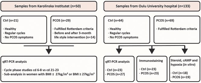
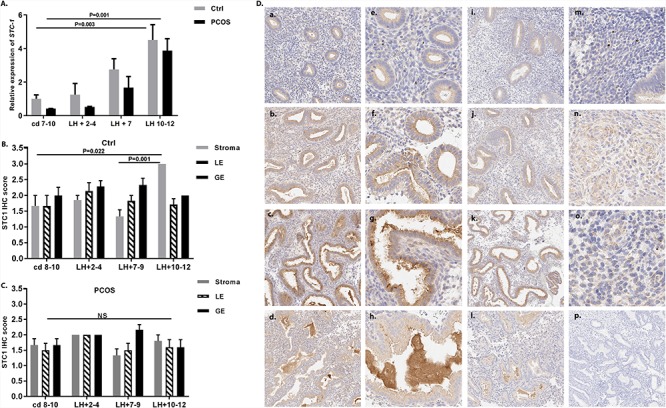
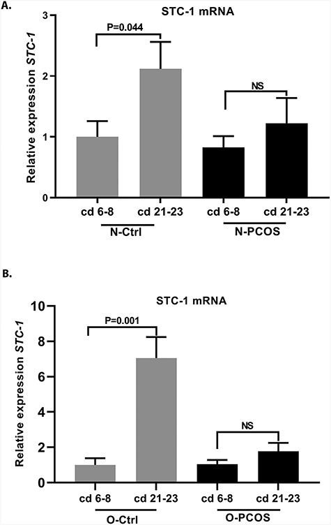
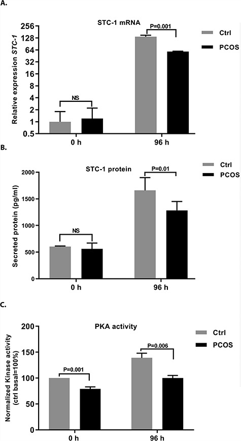
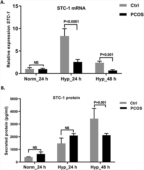
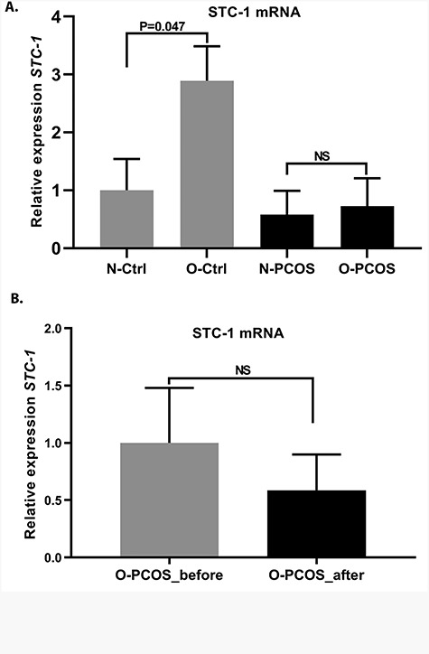
Stanniocalcin-1 (STC-1) is a pro-survival factor that protects tissues against stressors, such as hypoxia and inflammation. STC-1 is co-expressed with the endometrial receptivity markers, and recently endometrial STC-1 was reported to be dysregulated in endometriosis, a condition linked with endometrial progesterone resistance and inflammation. These features are also common in the endometrium in women with polycystic ovary syndrome (PCOS), the most common endocrine disorder in women. Given that women with PCOS present with subfertility, pregnancy complications, and increased risk for endometrial cancer, we investigated endometrial STC-1 expression in affected women. Endometrial biopsy samples were obtained from women with PCOS and controls, including samples from overweight/obese women with PCOS before and after a 3-month lifestyle intervention. A total of 98 PCOS and 85 control samples were used in immunohistochemistry, reverse-transcription polymerase chain reaction, or in vitro cell culture. STC-1 expression was analyzed at different cycle phases and in endometrial stromal cells (eSCs) after steroid hormone exposure. The eSCs were also challenged with 8-bromo-cAMP and hypoxia for STC-1 expression. The findings indicate that STC-1 expression is not steroid hormone mediated although secretory-phase STC-1 expression was blunted in PCOS. Lower expression seems to be related to attenuated STC-1 response to stressors in PCOS eSCs, shown as downregulation of protein kinase A activity. The 3-month lifestyle intervention did not restore STC-1 expression in PCOS endometrium. More studies are warranted to further elucidate the mechanisms behind the altered endometrial STC-1 expression and rescue mechanism in the PCOS endometrium.
Keywords: endometrial stromal cells; human endometrium; hypoxia; lifestyle intervention; polycystic ovary syndrome; primary cell culture; stanniocalcin-1; stress.
© The Author(s) 2019. Published by Oxford University Press on behalf of Society for the Study of Reproduction.
Figures
Similar articles
-
Mesenchymal stem/progenitors and other endometrial cell types from women with polycystic ovary syndrome (PCOS) display inflammatory and oncogenic potential.J Clin Endocrinol Metab. 2013 Sep;98(9):3765-75. doi: 10.1210/jc.2013-1923. Epub 2013 Jul 3. J Clin Endocrinol Metab. 2013. PMID: 23824412 Free PMC article.
-
Lifestyle intervention up-regulates gene and protein levels of molecules involved in insulin signaling in the endometrium of overweight/obese women with polycystic ovary syndrome.Hum Reprod. 2014 Jul;29(7):1526-35. doi: 10.1093/humrep/deu114. Epub 2014 May 19. Hum Reprod. 2014. PMID: 24842895
-
Endometrial stromal fibroblasts from women with polycystic ovary syndrome have impaired progesterone-mediated decidualization, aberrant cytokine profiles and promote enhanced immune cell migration in vitro.Hum Reprod. 2015 May;30(5):1203-15. doi: 10.1093/humrep/dev055. Epub 2015 Mar 6. Hum Reprod. 2015. PMID: 25750105 Free PMC article.
-
Endometrial function in women with polycystic ovary syndrome: a comprehensive review.Hum Reprod Update. 2021 Apr 21;27(3):584-618. doi: 10.1093/humupd/dmaa051. Hum Reprod Update. 2021. PMID: 33302299 Review.
-
Polycystic ovary syndrome: Endometrial markers.Best Pract Res Clin Obstet Gynaecol. 2016 Nov;37:66-79. doi: 10.1016/j.bpobgyn.2016.03.008. Epub 2016 Apr 1. Best Pract Res Clin Obstet Gynaecol. 2016. PMID: 27156350 Review.
Cited by
-
Implantation and Decidualization in PCOS: Unraveling the Complexities of Pregnancy.Int J Mol Sci. 2024 Jan 18;25(2):1203. doi: 10.3390/ijms25021203. Int J Mol Sci. 2024. PMID: 38256276 Free PMC article. Review.
-
Dissecting the shared genetic architecture between endometriosis and polycystic ovary syndrome.Front Endocrinol (Lausanne). 2024 Apr 29;15:1359236. doi: 10.3389/fendo.2024.1359236. eCollection 2024. Front Endocrinol (Lausanne). 2024. PMID: 38742190 Free PMC article.
-
Stanniocalcin-1 in the female reproductive system and pregnancy.Hum Reprod Update. 2021 Oct 18;27(6):1098-1114. doi: 10.1093/humupd/dmab028. Hum Reprod Update. 2021. PMID: 34432025 Free PMC article. Review.
-
The Disorders of Endometrial Receptivity in PCOS and Its Mechanisms.Reprod Sci. 2022 Sep;29(9):2465-2476. doi: 10.1007/s43032-021-00629-9. Epub 2021 May 27. Reprod Sci. 2022. PMID: 34046867 Review.
-
An Update on the Progress of Endometrial Receptivity in Women with Polycystic Ovary Syndrome.Reprod Sci. 2022 Aug;29(8):2136-2144. doi: 10.1007/s43032-021-00641-z. Epub 2021 Jun 2. Reprod Sci. 2022. PMID: 34076874 Review.
References
-
- Yoshiko Y, Aubin JE. Stanniocalcin 1 as a pleiotropic factor in mammals. Peptides 2004; 25:1663–1669. - PubMed
-
- Westberg JA, Serlachius M, Lankila P, Penkowa M, Hidalgo J, Andersson LC. Hypoxic preconditioning induces neuroprotective stanniocalcin-1 in brain via IL-6 signaling. Stroke 2007; 38:1025–1030. - PubMed
Publication types
MeSH terms
Substances
LinkOut - more resources
Full Text Sources
Medical
