

Lewy-related pathology exhibits two anatomically and genetically distinct progression patterns: a population-based study of Finns aged 85
- PMID: 31494694
- PMCID: PMC6800868
- DOI: 10.1007/s00401-019-02071-3
Lewy-related pathology exhibits two anatomically and genetically distinct progression patterns: a population-based study of Finns aged 85
Abstract
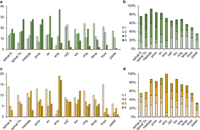
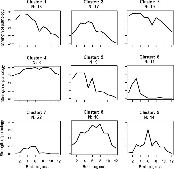
According to a generally accepted concept Lewy-related pathology (LRP) follows hierarchical caudo-rostral progression. LRP is also frequently present concomitantly with Alzheimer's disease (AD), and it has been hypothesized that AD-associated LRP forms a distinct type of α-synucleinopathy, where LRP originates in the amygdala. The frequency of distinct forms of LRP progression types has not been studied in a population-based setting. We investigated the distribution and progression of LRP and its relation to AD pathology and apolipoprotein (APOE) ε4 in a population-based sample of Finns aged over 85 years (N = 304). Samples from spinal cord to neocortical areas representing 11 anatomical sites without any hierarchical selection were analyzed immunohistochemically (α-synuclein antibody clone 5G4). LRP was present in 124 individuals (41%) and according to DLB Consortium guidelines 19 of them were categorized as brainstem, 10 amygdala-predominant, 41 limbic, and 43 diffuse neocortical type, whereas 11 could not be classified. To determine the LRP progression patterns, a systematic anatomical scoring was carried out by taking into account the densities of the semiquantitative LRP scores in each anatomic site. With this scoring 123 (99%) subjects could be classified into two progression pattern types: 67% showed caudo-rostral and 32% amygdala-based progression. The unsupervised statistical K-means cluster analysis was used as a supplementary test and supported the presence of two progression patterns and had a 90% overall concordance with the systematic anatomical scoring method. Severe Braak NFT stage, high CERAD score and APOE ε4 were significantly (all p < 0.00001) associated with amygdala-based, but not with caudo-rostral progression type (all p > 0.2). This population-based study demonstrates two distinct common LRP progression patterns in the very elderly population. The amygdala-based pattern was associated with APOE ε4 and AD pathology. The results confirm the previous progression hypotheses but also widen the concept of the AD-associated LRP.
Keywords: Aged, 80 and over; Alzheimer’s disease; Lewy body diseases; Lewy-related pathology; Population-based; α-Synuclein.
Conflict of interest statement
Pentti J. Tienari holds patent on C9orf72 in diagnostics and treatment of ALS/FTD and has made paid consultations to Roche, Biogen, Merck, Teva, Sanofi-Genzyme and Novartis. Other authors declare no conflict of interest.
Figures
References
Publication types
MeSH terms
Grants and funding
LinkOut - more resources
Full Text Sources
Medical
Molecular Biology Databases
Miscellaneous
