Review
. 2019 Oct;11(5):795-805.
doi: 10.1007/s12551-019-00584-5. Epub 2019 Sep 7.

Piezo1 mechanosensitive channels: what are they and why are they important

Affiliations
Review

Piezo1 mechanosensitive channels: what are they and why are they important

Pietro Ridone et al. Biophys Rev. 2019 Oct.

Abstract

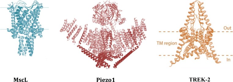
Mechanosensitive (MS) ion channels are integral membrane proteins which play a crucial role in fast signaling during mechanosensory transduction processes in living cells. They are ubiquitous and old in the evolutionary sense, given their presence in cells from all three kingdoms of life found on Earth, including bacterial, archaeal, and eukaryotic organisms. As molecular transducers of mechanical force, MS channels are activated by mechanical stimuli exerted on cellular membranes, upon which they rapidly and efficiently convert these stimuli into electrical, osmotic, and/or chemical intracellular signals. Most of what we know about the gating mechanisms of MS channels comes from the work carried out on bacterial channels. However, recent progress resulting from identification and structural information of eukaryotic K2P-type TREK and TRAAK as well as Piezo1 and Piezo2 MS channels has greatly contributed to our understanding of the common biophysical principles underlying the gating mechanism and evolutionary origins of these fascinating membrane proteins. Using Piezo1 channels as an example, we briefly describe in this review what we have learned about their biophysics, physiological functions, and potential roles in "mechanopathologies."

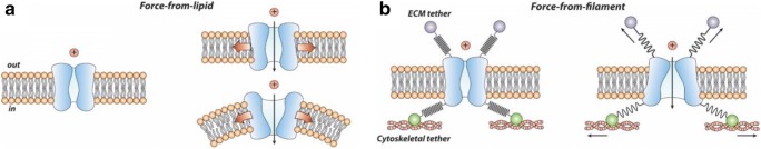
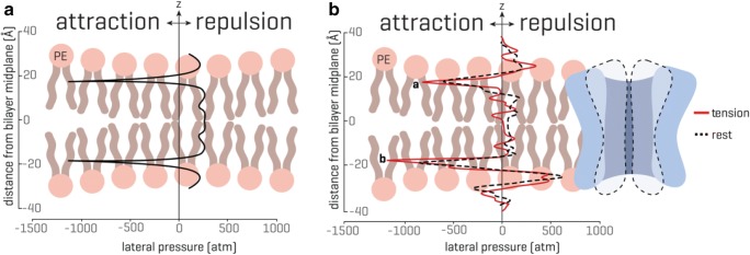
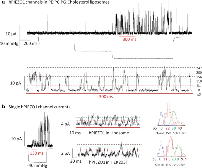
Keywords: Force-from-filament; Force-from-lipids; Lipid bilayer; Liposome reconstitution; Patch clamp; Transbilayer pressure profile.


Figures

Fig. 1
Fig. 1
Examples of truly mechanically activated ion channels. MscL, Piezo1, and TREK-2 are activated and controlled by mechanical force over their whole dynamic range from the fully closed to the fully open state. The 3D X-ray structure of MscL from Mycobacterium tuberculosis shows the channel homopentamer in the closed state (adapted from Chang et al. (1998)). The Piezo1 structure determined by cryo-EM shows the first low-resolution structure of the trimeric channel in its closed conformation (adopted from Ge et al. (2015)). The higher resolution structure has later been determined by several laboratories (Guo ; Zhao et al. ; Saotome et al. 2018). Crystal structure of the human TREK-2 channel, a member of the K2P family which forms dimers. Each monomer adds two-pore loops to the structure to end up with pseudo-tetrameric assembly (Dong et al. 2015). One of the structural characteristics of the truly mechanically gated channels is recently resolved lipid-binding domains, similar to the MscL N-terminus (Bavi et al. 2016). Such lipid-binding structural domains were identified in the MscS-like channels, K2P family of channels (TREK/TRAAK), and most recently Piezo1 ion channels (Bavi et al. 2017a, b)
Fig. 2
Fig. 2
Gating paradigms of MS channels. The two main generally accepted gating paradigms of MS channels are defined according to the cell membrane components (lipid bilayer (a) or ECM/CSK (b)) transmitting the activation force directly to MS channels
Fig. 3
Fig. 3
Transbilayer pressure profile. The transbilayer pressure profile is largely inhomogeneous across the bilayer thickness, which originates from the amphipathic nature of the lipid molecules and the presence of water. (A) An idealized symmetrical lipid bilayer. The transbilayer pressure profile shows characteristic negative peaks at the water-lipid interface (~ 1000 atm) and repulsive positive peaks (~ 300 atm) in the headgroup and tail region. The z indicates the bilayer thickness direction. (B) In the presence of a membrane protein, the pressure profile in a symmetrical lipid bilayer becomes noticeably asymmetric. Peak a and peak b represent the rise in the pressure profile at the lipid solvent interface (modified from Cox et al. ; for more details see also Bavi et al. 2016)
Fig. 4
Fig. 4
Cholesterol effect on Piezo1 clustering and channel kinetics. (A) Super-resolution microscopy shows the channels clustering together at the nanoscale (top). These clusters are dependent on the cholesterol content of the membrane (bottom). (B) Leaching cholesterol from the membrane changes sensitivity and gating kinetics. Under pressure application in a membrane patch (right control) the current decays as the channel inactivates. This behavior is lost if cholesterol is removed (top). Boltzmann distribution function showing dependence of Piezo1 currents on negative pressure (suction) applied to the cell-attached membrane patch of HEK293 cells. Piezo1 channels in cells treated with 5 mM MBCD exhibit reduced mechanosensitivity as indicated by a shallowed slope of the Boltzmann function (bottom left). The half activation pressure of Piezo1 channels increased significantly in membrane patches treated with MBCD (bottom right) (modified from Ridone et al. (2019))
Fig. 5
Fig. 5
Mechanical activation of purified human Piezo1 (hPiezo1) reconstituted in artificial liposomes made of PE:PC:PG:Cholesterol. (A) Representative inside-out patch clamp recording of hPIEZO1 at + 65 mV pipette potential. Inward currents produced in response to 10 mmHg incremental steps in suction. Bottom: inset shows the concerted activity of up to 5 hPIEZO1 channels and the observed conductance values (pS) are indicated on the right. (B) Mechanical activation of single PIEZO1 channel recorded in artificial liposomes and summarized in a representative histogram of conductance values on the right-hand side. Bottom: single hPIEZO1 activity in HEK293T cells transfected with the same construct used for protein purification and representative histogram of single channel events (liposome recording solution: 200 mM KCl, 40 mM MgCl2, 5 mM HEPES, pH 7.4. HEK293 cell recoding solution: 140 mM NaCl, 3 mM KCl, 1 mM MgCl2, 1 mM CaCl2, 10 mM glucose, 10 mM HEPES, pH 7.4) (unpublished results)

Similar articles

Cited by

References

    1. Albuisson J, Murthy SE, Bandell M, Coste B, Louis-Dit-Picard H, Mathur J, Feneant-Thibault M, Tertian G, De Jaureguiberry JP, Syfuss PY, Cahalan S, Garcon L, Toutain F, Simon Rohrlich P, Delaunay J, Picard V, Jeunemaitre X, Patapoutian A. Dehydrated hereditary stomatocytosis linked to gain-of-function mutations in mechanically activated PIEZO1 ion channels. Nat Commun. 2013;4:1884. - PMC - PubMed
    1. Bae C, Gnanasambandam R, Nicolai C, Sachs F, Gottlieb PA. Xerocytosis is caused by mutations that alter the kinetics of the mechanosensitive channel PIEZO1. Proc Natl Acad Sci U S A. 2013;110:E1162–E1168. - PMC - PubMed
    1. Barry RM, Gitai Z. Self-assembling enzymes and the origins of the cytoskeleton. Curr Opin Microbiol. 2011;14:704–711. - PMC - PubMed
    1. Bass RB, Strop P, Barclay M, Rees DC. Crystal structure of Escherichia coli MscS, a voltage-modulated and mechanosensitive channel. Science. 2002;298:1582–1587. - PubMed
    1. Bavi N, Nakayama Y, Bavi O, Cox CD, Qin QH, Martinac B. Biophysical implications of lipid bilayer rheometry for mechanosensitive channels. Proc Natl Acad Sci U S A. 2014;111:13864–13869. - PMC - PubMed