Review
. 2020 Jun;23(2):195-205.
doi: 10.1007/s40477-019-00405-1. Epub 2019 Sep 7.

Ultrasound findings in urogenital schistosomiasis: a pictorial essay

Affiliations
Review

Ultrasound findings in urogenital schistosomiasis: a pictorial essay

Diletta Cozzi et al. J Ultrasound. 2020 Jun.

Abstract

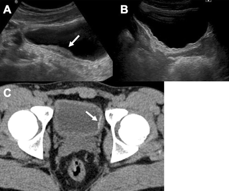
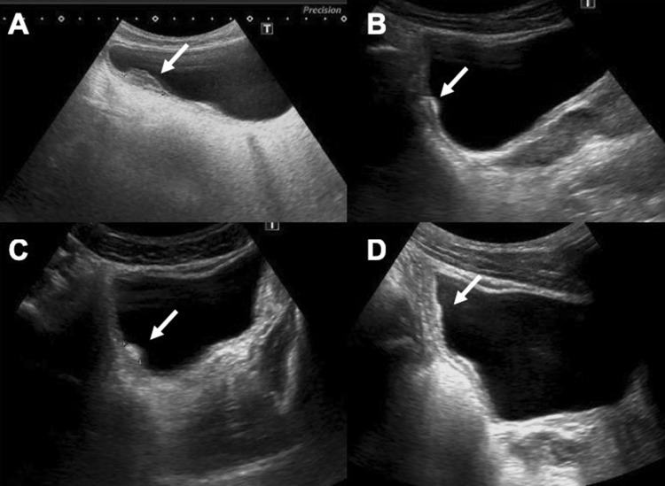
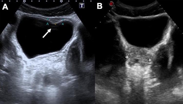
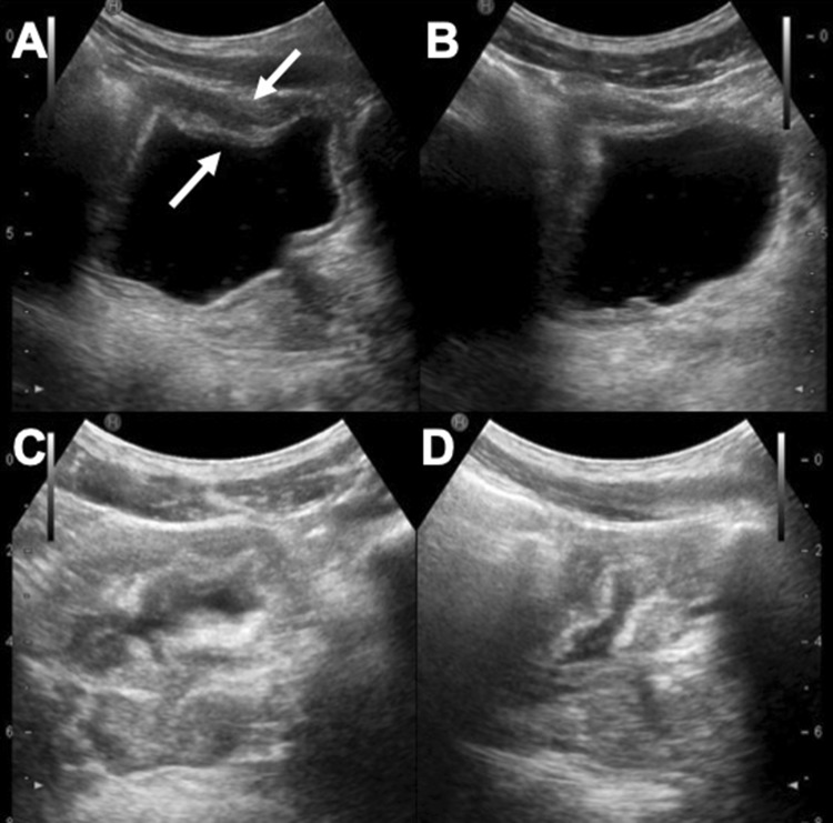
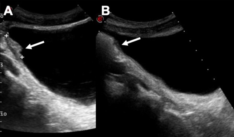
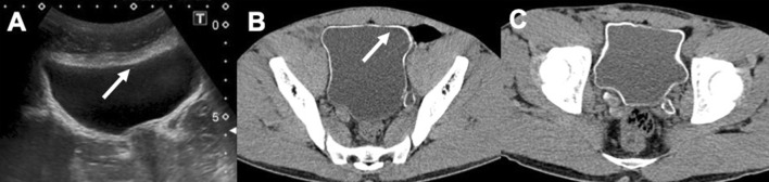
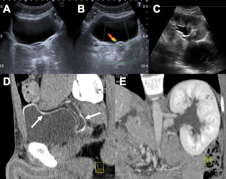
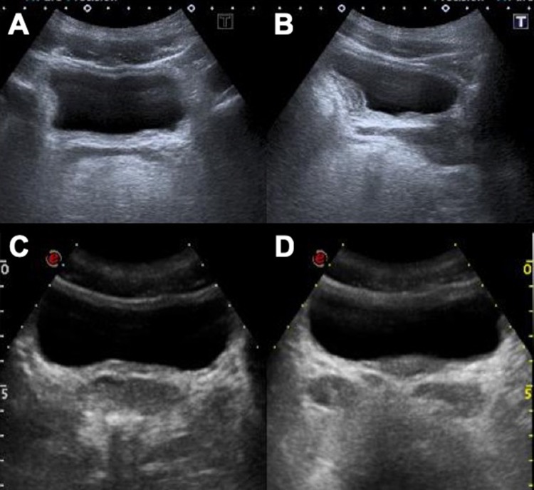
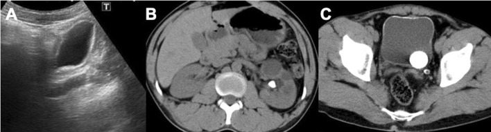
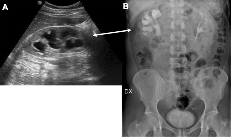
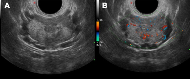
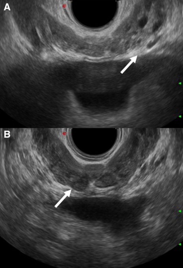
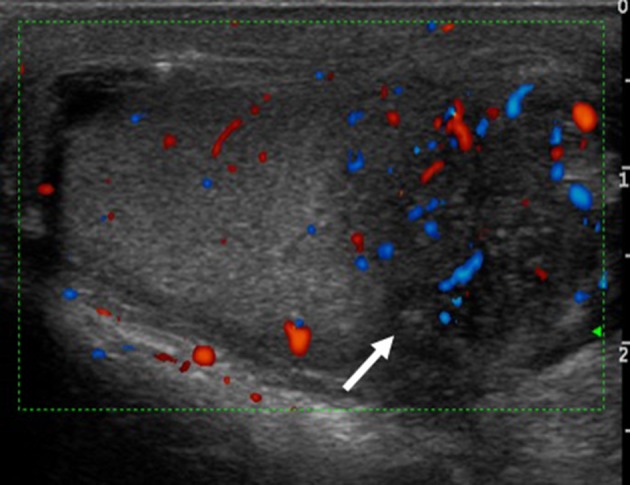
Urogenital schistosomiasis is a parasitic disease caused by S. haematobium which is endemic in tropical and sub-tropical areas but is increasingly diagnosed in temperate non-endemic countries due to migration and international travels. Early identification and treatment of the disease are fundamental to avoid associated severe sequelae such as bladder carcinoma, hydronephrosis leading to kidney failure and reproductive complications. Radiologic imaging, especially through ultrasound examination, has a fundamental role in the assessment of organ damage and follow-up after treatment. Imaging findings of urinary tract schistosomiasis are observed mainly in the ureters and bladder. The kidneys usually appear normal until a late stage of the disease.

Keywords: Bladder; Infection; Kidney; Schistosoma; Ultrasound.


Conflict of interest statement

The authors declare that they have no conflict of interest.

Figures

Fig. 1
Fig. 1
Vesical parietal thickening (arrow—a) and follow-up US control after 1 month of therapy (b). Image C shows a CT control after therapy. A small tiny calcification of the left bladder wall is still perceptible (arrow)
Fig. 2
Fig. 2
US scans in a, b show a focal nodular thickening of the bladder in a young Sub-Saharan man with haematuria (arrows). US follow-up after 3 months (c) and 5 months of therapy
Fig. 3
Fig. 3
Ultrasound suprapubic examination of a young man from Africa shows a nodular thickening of the bladder (arrow—a). The US follow-up control after 14 months does not show any parietal alteration (b)
Fig. 4
Fig. 4
Trilaminar aspect of the bladder walls (arrows, axial scan a; sagittal scan b) which remains even after vesical emptying (c, d)
Fig. 5
Fig. 5
Vesical parietal thickening (arrow—a) and follow-up US control after 4 months of therapy (b)
Fig. 6
Fig. 6
US and CT study of the bladder in a 24-year-old male from Senegal: the CT axial image demonstrates better the thin wall calcifications (arrows—a and b). CT follow-up after 20 months revealed a minimal increase in the thickness of parietal calcifications (c)
Fig. 7
Fig. 7
US images in a, b show a mild hyperechogenicity of the bladder wall associated with stages 1–2 hydronephrosis (c). CT sagittal reconstruction (d) demonstrates the bladder wall calcifications that extend also to the dilated ureter (arrow). Figure e is a para-coronal CT reconstruction that confirms the hydronephrosis
Fig. 8
Fig. 8
Diffuse parietal thickening of the bladder in a young Sub-Saharan man before (a, b) and after 14 months of treatment (c, d)
Fig. 9
Fig. 9
US scan in a shows a diffused hyperechogenicity of the bladder wall’s submucosa, confirmed in the CT study (b, c). Axial CT scan highlights also an early hydronephrosis with two big calculi
Fig. 10
Fig. 10
US imaging (a) shows stage 2 hydronephrosis of the right kidney, confirmed also with urography (b) in a young male patient from Africa
Fig. 11
Fig. 11
US study with transrectal approach demonstrates inflammatory areas (a) with hypervascularisation of the prostate gland (b)
Fig. 12
Fig. 12
Examples of prostate parenchymal calcifications (a), granulomatous flogosis (b) and pseudonodular inflammation of the gland (c) due to Schistosoma infection
Fig. 13
Fig. 13
a and b show ectasia of the seminal vesicles in two male patients affected by chronic schistosomiasis (arrows)
Fig. 14
Fig. 14
Chronic epididymitis in a young male patient: the arrow shows a mild increase in vascularization at the Doppler study. This finding is non-specific but falls within the possible manifestations of genitourinary schistosomiasis

References

    1. Colley DG, Bustinduy AL, Secor WE, King CH. Human schistosomiasis. Lancet. 2014;383:2253–2264. - PMC - PubMed
    1. Lingscheid T, Kurth F, Clerinx J, Marocco S, Trevino B, Schunk M, et al. Schistosomiasis in European travelers and migrants: analysis of 14 years TropNet Surveillance data. Am J Trop Med Hyg. 2017;97(2):567–574. - PMC - PubMed
    1. Chitsulo L, Engels D, Montresor A, Savioli L. The global status of schistosomiasis and its control. Acta Trop. 2000;77(1):41–51. - PMC - PubMed
    1. Buonfrate D, Gobbi F, Marchese V, Postiglione C, Badona-Monteiro G, Giorli G, et al. Extended screening for infectious diseases among newly-arrived asylum seekers from Africa and Asia, Verona province, Italy, April 2014 to June 2015. Eurosurveill. 2018 - PMC - PubMed
    1. Scarlata F, Giordano S, Romani A, Marasa L, Lipani G, Infurnari L, Titone L. Urinary schistosomiasis: remarks on a case. Infez Med. 2005;13(4):259–264. - PubMed

MeSH terms

LinkOut - more resources