. 2020 Jan;74(1):e22131.
doi: 10.1002/syn.22131. Epub 2019 Sep 23.

Synaptic vesicle fusion is modulated through feedback inhibition by dopamine auto-receptors

Affiliations

Synaptic vesicle fusion is modulated through feedback inhibition by dopamine auto-receptors

Rosaria Formisano et al. Synapse. 2020 Jan.

Abstract

Mechanisms of synaptic vesicular fusion and neurotransmitter clearance are highly controlled processes whose finely-tuned regulation is critical for neural function. This modulation has been suggested to involve pre-synaptic auto-receptors; however, their underlying mechanisms of action remain unclear. Previous studies with the well-defined C. elegans nervous system have used functional imaging to implicate acid sensing ion channels (ASIC-1) to describe synaptic vesicle fusion dynamics within its eight dopaminergic neurons. Implementing a similar imaging approach with a pH-sensitive fluorescent reporter and fluorescence resonance after photobleaching (FRAP), we analyzed dynamic imaging data collected from individual synaptic termini in live animals. We present evidence that constitutive fusion of neurotransmitter vesicles on dopaminergic synaptic termini is modulated through DOP-2 auto-receptors via a negative feedback loop. Integrating our previous results showing the role of ASIC-1 in a positive feedback loop, we also put forth an updated model for synaptic vesicle fusion in which, along with DAT-1 and ASIC-1, the dopamine auto-receptor DOP-2 lies at a modulatory hub at dopaminergic synapses. Our findings are of potential broader significance as similar mechanisms are likely to be used by auto-receptors for other small molecule neurotransmitters across species.

Keywords: C. elegans; FRAP; auto-receptor; dopamine; neurotransmitter; synaptic modulation.


Figures

FIGURE 1
FIGURE 1
Fluorescence recovery after photobleaching (FRAP) at dopaminergic synapses labeled with SNB-1::SEpHluorin is significantly increased in animals carrying a lesion in the dop-2 gene (red), compared with wild-type N2 animals (gray). FRAP recovery rate in animals carrying a lesion in the asic-1 gene (encodes an acid sensing ion channel) is markedly reduced (green) as also reported previously (Voglis & Tavernarakis, 2008). While ASIC-1 has been proposed to facilitate a positive feedback loop that reinforces constitutive dopamine release results presented in the current study indicate the existence of a distinct negative feedback mediated through DOP-2 auto-receptors. Given the biphasic nature of recovery, the data were fitted to an exponential recovery with two time constants (solid lines; see Methods). Setting complete FRAP recovery for wild-type N2 animals (M = 100%), we calculated the two time constants to be τ1 = 3.8 s, τ2 = 274 s and f = 0.27. The dop-2 deletion animals show a higher recovery, M = 150%, while their time scale of recovery is very similar to WT (τ1 = 3.8 s, τ2 = 274 s, f = 0.2). The asic-1 deletion mutants display both lesser recover and slower recovery dynamics (M = 45%, τ1 = 2.3 s, τ2 = 192 s, f = 0.25). Sample size of n = 30 synapses for wild-type N2 animals, n = 28 synapses for dop-2 deletion mutant, and n = 28 synapses for asic-1 deletion mutant
FIGURE 2
FIGURE 2
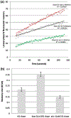
Slow recovery phase of wild-type animals and both dop-2 and asic-1 deletion mutants display distinct slopes as determined by linear regression (a). As compared to wild type, a significantly higher recovery for dop-2 deletion mutants and lower recovery for asic-1 deletions are observed (b). Error bars shown in the bar chart denote 95% confidence intervals for the slopes estimated using bootstrapping (p < 10–4)
FIGURE 3
FIGURE 3
An updated model for modulation of synaptic vesicle release at dopaminergic neuron synapses. Fusion of acidified neurotransmitter vesicles with the synaptic membrane causes release of dopamine and H+ ions. Dopamine activates a feedback loop through auto-receptors (DOP-2), and possibly through the membrane transporter (DAT-1), while ASIC-1 functions to facilitate a positive feedback loop that reinforces dopamine release in response to a local pH drop in the synaptic cleft. Thus, DOP-2, DAT-1 and ASIC-1 form a modulatory hub responsible for fine-tuning synaptic dopamine levels

Similar articles

Cited by

References

    1. Beaulieu JM, & Gainetdinov RR (2011). The physiology, signaling, and pharmacology of dopamine receptors. Pharmacology Reviews, 63(1), 182–217. 10.1124/pr.110.002642 - DOI - PubMed
    1. Bermingham DP, Hardaway JA, Snarrenberg CL, Robinson SB, Folkes OM, Salimando GJ, … Blakely RD (2016). Acute blockade of the Caenorhabditis elegans dopamine transporter DAT-1 by the mammalian norepinephrine transporter inhibitor nisoxetine reveals the influence of genetic modifications of dopamine signaling in vivo. Neurochemistry International, 98, 122–128. - PMC - PubMed
    1. Chase DL, & Koelle MR (2007). Biogenic amine neurotransmitters in C. elegans. WormBook, the online review of C. elegans biology. WormBook. 10.1895/wormbook.1.132.1. - DOI - PMC - PubMed
    1. Duerr JS, Frisby DL, Gaskin J, Duke A, Asermely K, Huddleston D, … Rand JB (1999). The cat-1 gene of Caenorhabditis elegans encodes a vesicular monoamine transporter required for specific monoamine-dependent behaviors. Journal of Neuroscience, 19, 72–84. - PMC - PubMed
    1. Ford CP (2014). The role of D2-autoreceptors in regulating dopamine neuron activity and transmission. Neuroscience, 282, 13–22. 10.1016/j.neuroscience.2014.01.025 - DOI - PMC - PubMed

Publication types

MeSH terms