. 2019 Oct 11;125(9):834-846.
doi: 10.1161/CIRCRESAHA.119.314794. Epub 2019 Sep 9.

Yin Yang 1 Suppresses Dilated Cardiomyopathy and Cardiac Fibrosis Through Regulation of Bmp7 and Ctgf

Affiliations

Yin Yang 1 Suppresses Dilated Cardiomyopathy and Cardiac Fibrosis Through Regulation of Bmp7 and Ctgf

Chia Yee Tan et al. Circ Res. .

Abstract

Rationale: Pathogenic variations in the lamin gene (LMNA) cause familial dilated cardiomyopathy (DCM). LMNA insufficiency caused by LMNA pathogenic variants is believed to be the basic mechanism underpinning LMNA-related DCM.

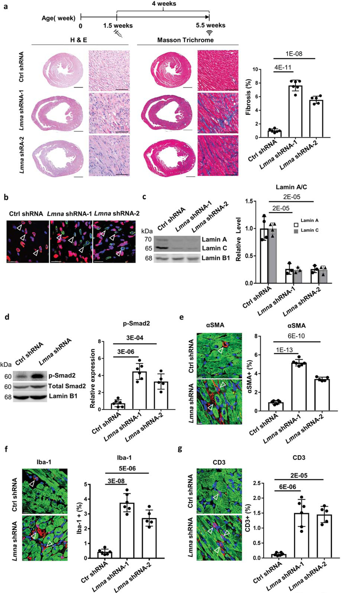
Objective: To assess whether silencing of cardiac Lmna causes DCM and investigate the role of Yin Yang 1 (Yy1) in suppressing Lmna DCM.

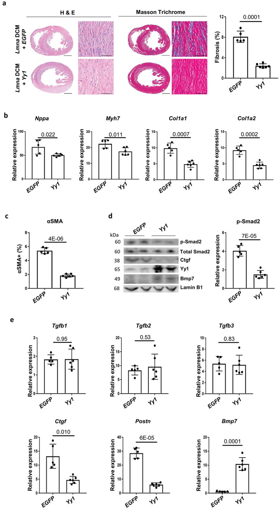
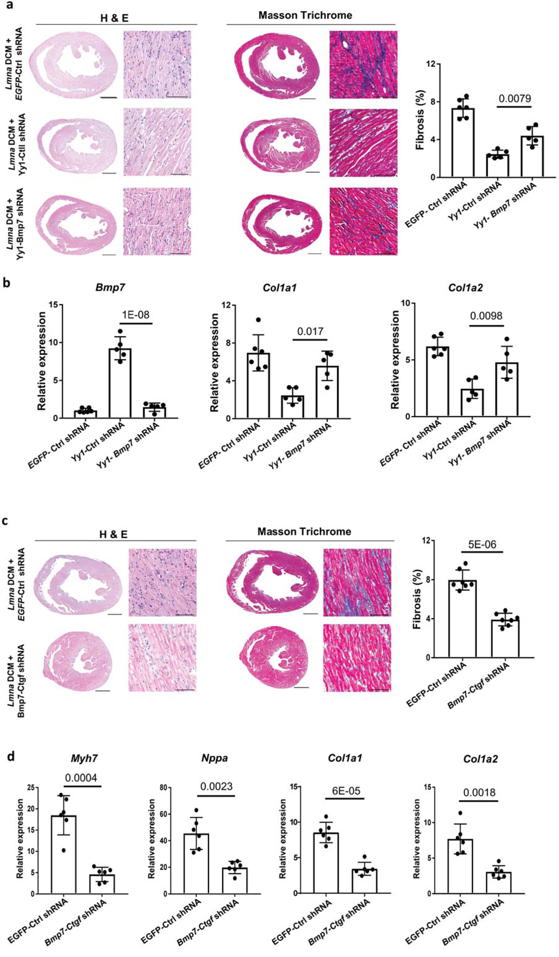
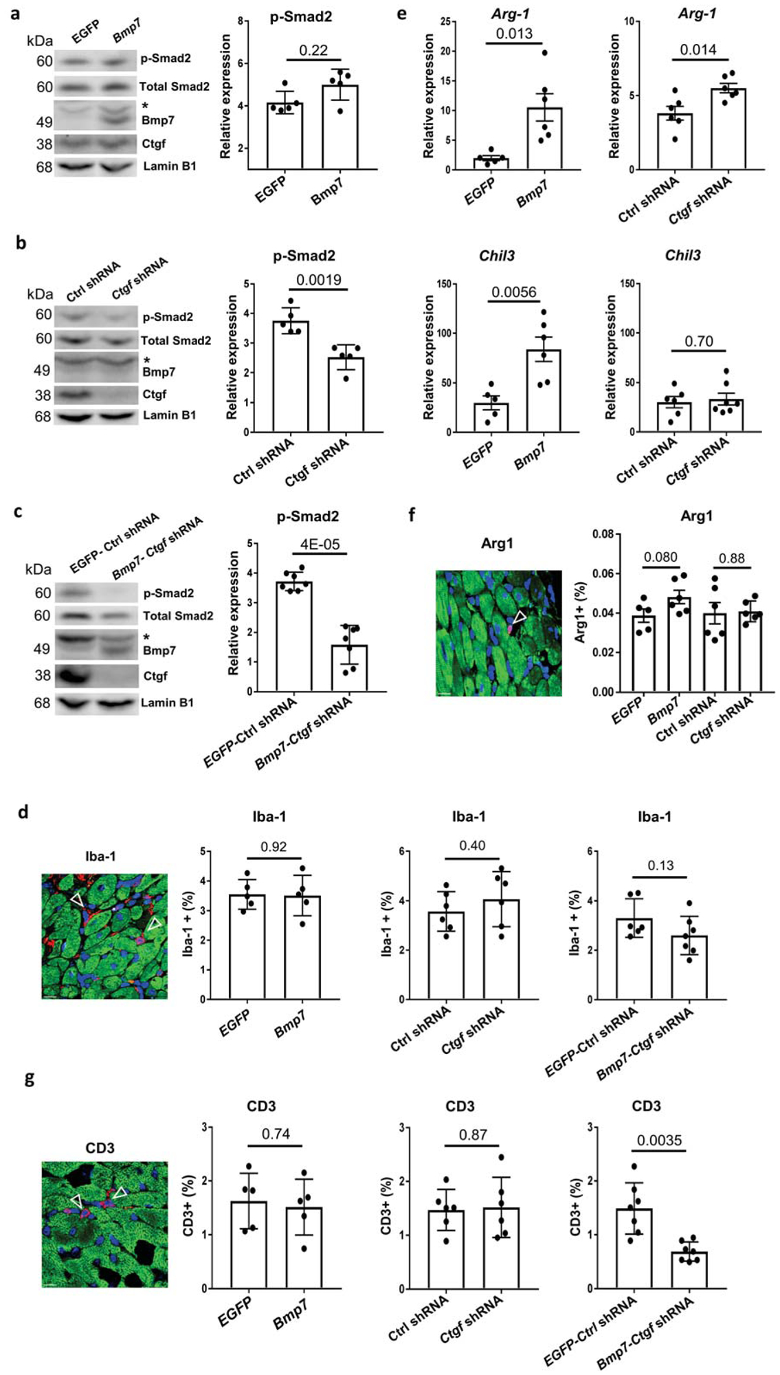
Methods and results: We developed a Lmna DCM mouse model induced by cardiac-specific Lmna short hairpin RNA. Silencing of cardiac Lmna induced DCM with associated cardiac fibrosis and inflammation. We demonstrated that upregulation of Yy1 suppressed Lmna DCM and cardiac fibrosis by inducing Bmp7 expression and preventing upregulation of Ctgf. Knockdown of upregulated Bmp7 attenuated the suppressive effect of Yy1 on DCM and cardiac fibrosis. However, upregulation of Bmp7 alone was not sufficient to suppress DCM and cardiac fibrosis. Importantly, upregulation of Bmp7 together with Ctgf silencing significantly suppressed DCM and cardiac fibrosis. Mechanistically, upregulation of Yy1 regulated Bmp7 and Ctgf reporter activities and modulated Bmp7 and Ctgf gene expression in cardiomyocytes. Downregulation of Ctgf inhibited TGF-β (transforming growth factor-β)/Smad signaling in DCM hearts. Regulation of both Bmp7 and Ctgf further suppressed TGFβ/Smad signaling. In addition, co-modulation of Bmp7 and Ctgf reduced CD3+ T cell numbers in DCM hearts.

Conclusions: Our findings demonstrate that upregulation of Yy1 or co-modulation of Bmp7 and Ctgf offer novel therapeutic strategies for the treatment of DCM caused by LMNA insufficiency.

Keywords: cardiomyopathies; fibrosis; genetic therapy; inflammation; transcription factors; upregulation.


Figures

Figure 1.
Figure 1.. Cardiac specific Lmna shRNA induces DCM in mice.
(a) Experimental timeline showing timepoints of virus injection and echocardiogram. Cardiac performance was assessed by echocardiogram at 5.5 week old. H&E and Masson Trichrome (MT) staining was performed on paraffin heart sections taken 4 weeks after control shRNA and Lmna shRNA transduction. Quantification of myocardial fibrosis of MT sections is shown, virus dose, 2.0E+13 vg/kg, n ≥ 5. Scale bars: 1000 μm for complete heart images; 100 μm for enlarged images. (b) Paraffin heart section immunostained for Lamin A/C (red), PCM1 (green) and DAPI (blue) in mice transduced with control shRNA and Lmna shRNA, scale bar = 100 μm. (c, d) Western blot and quantitative analysis of Lamin A/C protein levels in isolated cardiomyocytes (c) and phospho-Smad2 protein levels in mouse heart tissues (d), n ≥ 3. Data were normalized to Lamin B1. (e-g) Paraffin heart sections (left) and quantifications (right) of (e) αSMA (red), (f) Iba-1 (red) and (g) CD3 (red), cTnI (green) and DAPI (blue) positive cells in mice transduced with control shRNA (top) and Lmna shRNA (bottom), n ≥ 5, scale bar = 50 μm.
Figure 2.
Figure 2.. Yy1 suppresses Lmna DCM and cardiac fibrosis
(a) H&E and MT staining of paraffin heart sections of Lmna DCM mice treated with EGFP or Yy1. Quantification of myocardial fibrosis of MT sections is shown, n ≥ 5. Scale bars: 1000 μm for complete heart images; 100 μm for enlarged images. (b) Quantitative real-time PCR analyses of Nppa, Myh7, Col1a1 and Col1a2 expressions in EGFP and Yy1 treated groups. Mouse hearts were harvested 4 weeks after transduction, n ≥ 5. (c) Quantification of αSMA positive cells in paraffin heart sections from Lmna DCM mice treated with EGFP and Yy1, n ≥ 5. (d) Western blot and quantitative analysis of phospho-Smad2 (p-Smad2) protein levels in mouse heart tissue of Lmna DCM mice treated with EGFP and Yy1, n ≥ 5. Data were normalized to Lamin B1. (e) Quantitative real-time PCR analyses of Tgfb1, Tgfb2, Tgfb3, Ctgf, Postn and Bmp7 in Lmna DCM mice treated with EGFP and Yy1, n ≥ 5.
Figure 3.
Figure 3.. Co-regulation of Bmp7 and Ctgf suppresses Lmna DCM and cardiac fibrosis
(a and c) H&E (left panel) and MT (middle panel) staining of paraffin heart sections of Lmna DCM mice 4 weeks after (a) EGFP-Ctrl shRNA, Yy1-Ctrl shRNA or Yy1-Bmp7 shRNA transduction and (c) EGFP-Ctrl shRNA or Bmp7-Ctgf shRNA transduction. Quantification of myocardial fibrosis (right panel) from MT-stained sections, n ≥ 6. Scale bars: 1000 μm for complete heart images; 100 μm for enlarged images. (b) Quantitative real-time PCR analyses of Bmp7, Col1a1 and Col1a2 expression in Lmna DCM groups treated with EGFP-Ctrl shRNA, Yy1-Ctrl shRNA or Yy1-Bmp7 shRNA, n ≥ 5. (d) Quantitative real-time PCR analyses of Myh7, Nppa, Col1a1 and Col1a2 expressions in Lmna DCM groups treated with EGFP-Ctrl shRNA or Bmp7-Ctgf shRNA groups, n ≥ 6.
Figure 4.
Figure 4.. Bmp7 and Ctgf regulate cardiac inflammation and TGFβ signaling
(a-c) Western blot (left panel) and quantitative analysis (right panel) of phospho-Smad2 (p-Smad2) protein levels in mouse heart tissue of Lmna DCM groups treated with (a) EGFP or Bmp7; (b) Ctrl shRNA or Ctgf shRNA or (c) EGFP Ctrl shRNA or Bmp7-Ctgf shRNA, n ≥ 5. Data were normalized to Lamin B1.* indicates the non-specific band. (d, f and g) Paraffin heart section staining (left panel) and quantifications (right pane) of (d) Iba-1 (red), (f) Arginase 1 (red) and (g) CD3 (red), cTnI (green) and DAPI (blue) positive cells in Lmna DCM groups treated with EGFP, Bmp7, Ctrl shRNA, Ctgf shRNA, EGFP-Ctrl shRNA or Bmp7-Ctgf shRNA, scale bar = 50 μm, n ≥5. (e) Quantitative real-time PCR analyses of Arg-1 and Chl3 expression in Lmna DCM groups treated with EGFP, Bmp7, Ctrl shRNA, Ctgf shRNA, n ≥ 5.

References

    1. Weintraub RG, Semsarian C, Macdonald P. Dilated cardiomyopathy. Lancet. 2017;390:400–414 - PubMed
    1. Hershberger RE, Hedges DJ, Morales A. Dilated cardiomyopathy: The complexity of a diverse genetic architecture. Nat Rev Cardiol. 2013;10:531–547 - PubMed
    1. McNally EM, Golbus JR, Puckelwartz MJ. Genetic mutations and mechanisms in dilated cardiomyopathy. J Clin Invest. 2013;123:19–26 - PMC - PubMed
    1. Lu JT, Muchir A, Nagy PL, Worman HJ. Lmna cardiomyopathy: Cell biology and genetics meet clinical medicine. Dis Model Mech. 2011;4:562–568 - PMC - PubMed
    1. Sullivan T, Escalante-Alcalde D, Bhatt H, Anver M, Bhat N, Nagashima K, Stewart CL, Burke B. Loss of a-type lamin expression compromises nuclear envelope integrity leading to muscular dystrophy. J Cell Biol. 1999;147:913–920 - PMC - PubMed

Publication types

MeSH terms