. 2019;8(1):1300-1313.
doi: 10.1080/22221751.2019.1659552.

Marburg virus regulates the IRE1/XBP1-dependent unfolded protein response to ensure efficient viral replication

Affiliations

Marburg virus regulates the IRE1/XBP1-dependent unfolded protein response to ensure efficient viral replication

Cornelius Rohde et al. Emerg Microbes Infect. 2019.

Abstract

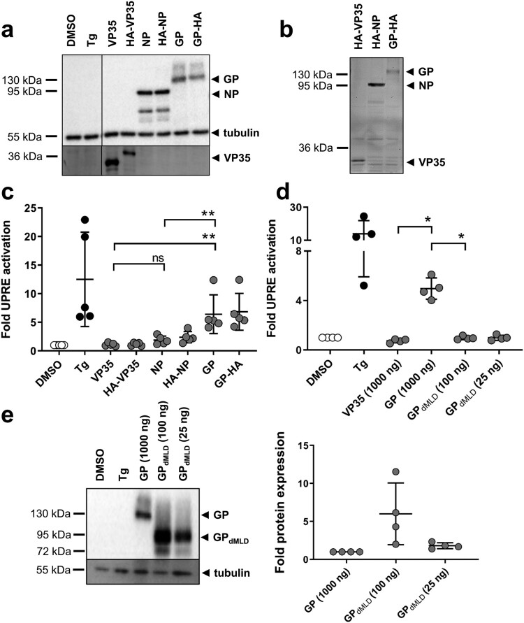
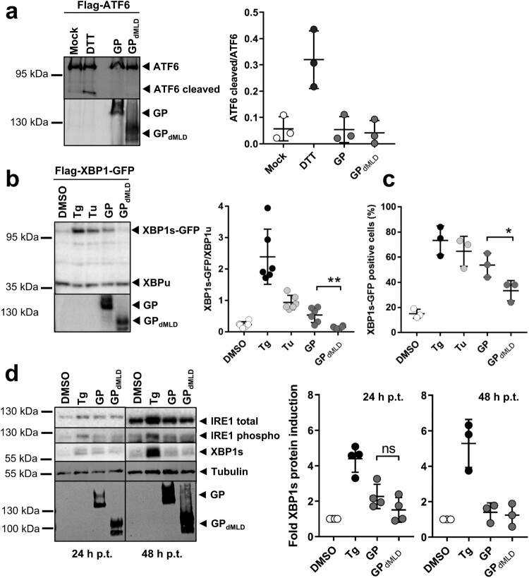
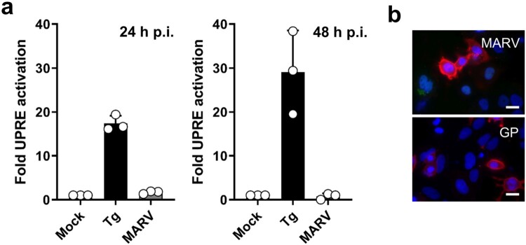
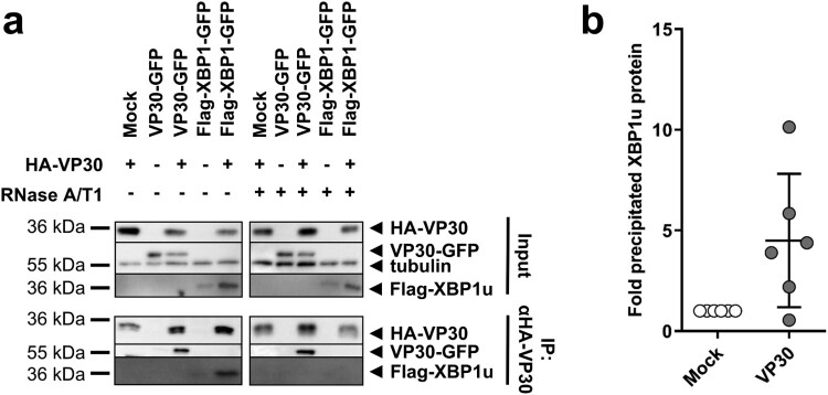
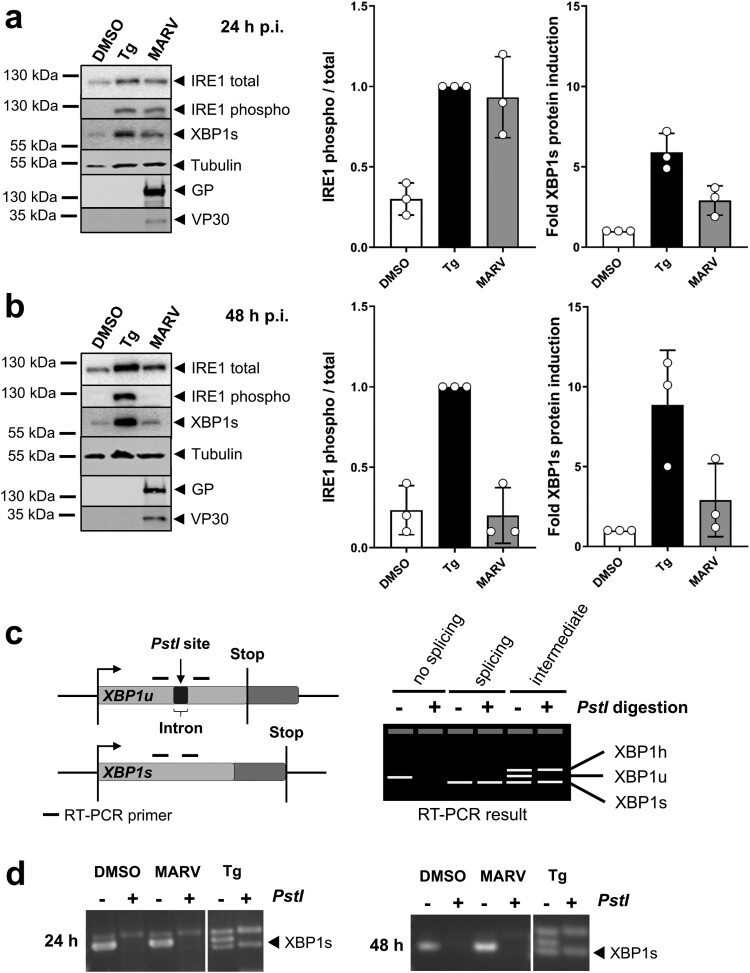
Viruses regulate cellular signalling pathways to ensure optimal viral replication. During Marburg virus (MARV) infection, large quantities of the viral glycoprotein GP are produced in the ER; this may result in the activation of the unfolded protein response (UPR). The most conserved pathway to trigger UPR is initiated by IRE1. Activation of IRE1 results in auto-phosphorylation, splicing of the XBP1 mRNA and translation of the XBP1s protein. XBP1s binds cis-acting UPR elements (UPRE) which leads to the enhanced expression of genes which should restore ER homeostasis. XBP1u protein is translated, if IRE1 is not activated. Here we show that ectopic expression of MARV GP activated the IRE1-XBP1 axis of UPR as monitored by UPRE luciferase assays. However, while at 24 h of infection with MARV IRE1 was phosphorylated, expression of XBP1s was only slightly enhanced and UPRE activity was not detected. The IRE1-XBP1 axis was not active at 48 h p.i. Co-expression studies of MARV proteins demonstrated that the MARV protein VP30 suppressed UPRE activation. Co-immunoprecipitation analyses revealed an RNA-dependent interaction of VP30 with XBP1u. Knock-out of IRE1 supported MARV infection at late time points. Taken together, these results suggest that efficient MARV propagation requires specific regulation of IRE1 activity.

Keywords: ER stress; GP; IRE1; Marburg virus; VP30; XBP1.


Conflict of interest statement

No potential conflict of interest was reported by the authors.

Figures

Figure 1.
Figure 1.
MARV GP activates the unfolded protein response element. (a) HuH7 cells were transfected with plasmids encoding firefly luciferase under the control of an UPRE promoter, with pGL4.73, which encodes Renilla luciferase, and with plasmids encoding NP, VP35 or GP. HuH7 cells transfected with the empty vector were treated with vehicle (DMSO) or with Tg. The cells were lysed at 48 h post transfection (p.t.), and equal amounts of the cell lysates were subjected to Western blotting using monoclonal antibodies against GP, NP and tubulin and polyclonal anti-VP35 serum. The experiment was performed five times; the results of one representative experiment are shown. (b) Equal amounts of cell lysates were subjected to SDS-PAGE, and the gels were subsequently incubated with anti-HA antibodies to detect HA-tagged viral proteins. (c) Cell lysates were analysed using luciferase assays. Firefly luciferase activity was normalized to Renilla activity, and the fold activation in comparison to the DMSO control (set to 1) was calculated. The experiment was performed five times. Statistical analysis was performed for wildtype proteins. (d) HuH7 cells were treated and transfected as described in (a) except that the amount of GPdMLD-expressing plasmid used for transfection was reduced (25 or 100 ng). The total amount of transfected plasmid was kept constant by the addition of empty vector. The experiment was performed four times. (e) Cell lysates were subjected to Western blotting using monoclonal antibodies to detect MARV GP and tubulin. Protein amount was quantified in each of the four independent experiments shown in d. Each circle represents a sample from an individual experiment, data are shown as the means ± SD.
Figure 2.
Figure 2.
MARV GP activates UPR in an IRE1/XBP1-dependent manner. (a) HuH7 cells transfected with plasmids encoding Flag-ATF6 and GP or GPdMLD (1 µg each) were lysed 48 h p.t. and analysed by Western blotting using an anti-Flag mouse monoclonal antibody and an Alexa680-conjugated anti-mouse antibody to detect full-length and cleaved (active) ATF6. MARV-specific goat serum and an IRdye800-conjugated anti-goat antibody were used to detect the viral proteins. Incubation of cells with 1 mM DTT for 30 min served as a positive control. Detection and quantification were performed using an Odyssey imaging system. The ratio of cleaved ATF6 protein to full-length ATF6 protein was calculated. The experiment was performed three times. (b) HuH7 cells were transfected with plasmids encoding Flag-XBP1-GFP, GP (1 µg), GPdMLD (25 ng) or empty vector (DMSO, Tg, Tu). The total amount of transfected plasmid (2 µg in total) was kept constant by the addition of empty vector. XBP1 splicing was induced by 5 nM Tg or 300 nM Tu for 16 h. The cells were lysed at 48 h p.t. and analysed by Western blotting using monoclonal antibodies against the Flag-tag and GP and peroxidase-coupled secondary antibodies. XBP1s and XBP1u were quantified using the ChemiDoc imaging system, and the ratios of these proteins were calculated. The experiment was performed six times. (c) HuH7 cells that had been treated and transfected as explained in b were fixed 48 h p.t. and subjected to immunofluorescence analysis. DMSO, Tg and Tu: HuH7 cells were transfected with an mCherry-expressing plasmid instead of with empty vector and were treated as indicated in b. Viral proteins were stained using monoclonal protein-specific and fluorescently labelled secondary antibodies. XBP1s-GFP positive nuclei were counted in cells expressing the viral protein or mCherry in three independent experiments. The percentage of XBP1s-GFP positive nuclei is shown. Each circle represents the result from an individual experiment, data are shown as the means ± SD. (d) HuH7 cells were transfected with plasmids encoding GP (1 µg), GPdMLD (200 ng) or mCherry (DMSO, Tg). The total amount of transfected plasmid (2 µg in total) was kept constant by the addition of mCherry plasmid. Cells were lysed at 24 and 48 h p.t. and subjected to Western blot analysis to detect endogenous IRE1 and XBP1s proteins using protein-specific antibodies detected by POD-coupled secondary antibodies. 24 and 48 h samples were analysed in parallel on the same blot afterwards tubulin and MARV GP were detected. XBP1s levels were quantified and presented as relative values to DMSO-treated cells (set to 1). The experiments were performed four (24 h) or three (48 h) times. Each circle represents a sample from an individual experiment, data are shown as the means ± SD.
Figure 3.
Figure 3.
MARV infection does not induce UPRE. (a) The UPRE firefly luciferase assay was performed as described in the legend to Figure 1. At 24 h p.t. the cells were infected with MARV at a MOI of 1. Tg (300 nM for 24 h) was used to activate the UPRE reporter. The cells were lysed at 24 or 48 h post infection (p.i.) and analysed using the luciferase assay. The experiments were performed three times. Each circle represents a sample from an individual experiment, data are shown as the means ± SD. (b) HuH7 cells were infected with MARV (see above) or transfected with plasmids encoding GP as described in the legend to Figure 1. The cells were fixed after 48 h and subjected to IFA using a monoclonal antibody against GP. The photomicrographs were obtained using the same exposure times. DAPI staining labels cell nuclei. Scale bar = 25 µm.
Figure 4.
Figure 4.
VP30 reduces GP- and Tg-induced UPRE-dependent signalling. (a) HuH7 cells were transfected with plasmids encoding the indicated MARV proteins and the UPRE-specific luciferase reporter plasmids as described in the legend to Figure 1. To express viral proteins, 0.5 µg of each plasmid was used in the transfection. In the iVLP setting, which involved the use of a combination of plasmids encoding all MARV proteins, the plasmid amounts used in transfection were as described by Wenigenrath et al. [25]. The experiment was repeated 4 times. (b) Equal amounts of lysates of transfected HuH7 cells were subjected to Western blot analysis using monoclonal antibodies. NP, GP, VP30, and tubulin were detected simultaneously; VP40 was stained afterwards on the same blot. The asterisk indicates remaining VP30 staining; irrelevant lines have been removed. (c) VP30-dependent reduction of Tg-induced UPR. Tg (5 nM) was used to induce UPRE-dependent reporter gene expression in VP30-, VP35-, and GP-expressing cells that had been transfected as described in the legend to Figure 1. The experiment was repeated 4 times. (d) Western blot analysis of cell lysates obtained from c. VP35 was stained with a polyclonal antibody against VP35; GP, VP30, and tubulin were detected afterwards on the same blot using monoclonal antibodies. Each circle represents a sample from an individual experiment, data are shown as the means ± SD. (e) To analyse UPRE-dependent luciferase activity, HAP1 cells (wt, shown in yellow) or HAP1 IRE1 KO cells (shown in pink) were transfected, treated and harvested as described for HuH7 cells. To restore IRE1 signalling in KO cells, the KO cells were transfected with a plasmid encoding IRE1 (100 ng); The cells were treated either with vehicle (DMSO) or with 5 nM Tg for 16 h. The experiments were performed three times. Each circle represents a sample from an individual experiment, data are shown as the means ± SD.
Figure 5.
Figure 5.
VP30 co-precipitates XBP1u protein in the presence of RNA. (a) HuH7 cells were transfected with plasmids expressing Flag-XBP1, VP30-GFP and HA-VP30. The cells were lysed 48 h p.t. and expression of ectopically expressed proteins was checked (input). The remaining lysate was subjected to co-immunoprecipitation analysis using anti-HA agarose according to Biedenkopf et al. [34]. (b) The amount of XBP1u precipitated in the presence of VP30 was compared with the amount precipitated in the absence of VP30 (set to 1). The amount of precipitated XBP1u protein was normalized to the expressed XBP1u (input) according to the tubulin content of the lysate. The experiment was performed six times. Each circle represents a sample from an individual experiment, data are shown as the means ± SD.
Figure 6.
Figure 6.
IRE1-dependent signalling during MARV infection. (a, b) HuH7 cells were infected with MARV at a MOI of 1. Cells were lysed at 24 h (a) and 48 h p.i. (b) and subjected to Western blot analysis to detect endogenous IRE1 (total and phosphorylated) and XBP1s proteins as explained in the legend to Figure 3. Total and phosphorylated IRE1 was quantified in each sample, compared to each other and set in relation to Tg-treated samples (set to 1). XBP1s levels were quantified and presented as relative values to DMSO-treated cells (set to 1). The experiments were performed three times. Each circle represents a sample from an individual experiment, data are shown as the means ± SD. (c) Scheme of XBP1-specific mRNAs and RT-PCR results. If there is no IRE1 activity, XBP1u mRNA is not spliced by IRE1; the PstI restriction site is available and the PCR product can be digested. Under conditions of IRE1 activation, XBP1u is spliced; PstI restriction site is lost and the PCR product cannot be digested by the enzyme. Intermediate phenotype: XBP1u is partially spliced; As published by others [39] we detect that XBP1u and XBP1s form a hybrid (XBP1 h, confirmed by sequencing) that is visible in the agarose gel and resistant to digestion. (d) XBP1-specific RT-PCR of RNA derived from HuH7 cells infected with MARV at a MOI of 1 for the indicated times. XBP1 splicing was induced using 5 nM Tg.
Figure 7.
Figure 7.
MARV propagation is affected by IRE1. (a) HAP1 cells (wt, shown in yellow) and HAP1 IRE1 KO cells (shown in pink) (6 × 105 cells) were infected with MARV at a MOI of 0.1; the cell culture supernatants were collected after 24, 48, 72 and 144 h p.i. and analysed for the presence of infectious MARV by TCID50 assays.

References

    1. Iranpour M, Moghadam AR, Yazdi M, et al. Apoptosis, autophagy and unfolded protein response pathways in arbovirus replication and pathogenesis. Expert Rev Mol Med. 2016;18:e1. DOI: 10.1017/erm.2015.19. - DOI - PMC - PubMed
    1. Schulz KS, Mossman KL.. Viral evasion strategies in type I IFN signaling – a summary of recent developments. Front Immunol. 2016;7:498. DOI: 10.3389/fimmu.2016.00498. - DOI - PMC - PubMed
    1. Ron D, Walter P.. Signal integration in the endoplasmic reticulum unfolded protein response. Nat Rev Mol Cell Biol. 2007;8:519–529. DOI: 10.1038/nrm2199. - DOI - PubMed
    1. Shore GC, Papa FR, Oakes SA.. Signaling cell death from the endoplasmic reticulum stress response. Curr Opin Cell Biol. 2011;23:143–149. DOI: 10.1016/j.ceb.2010.11.003. - DOI - PMC - PubMed
    1. Sano R, Reed JC.. ER stress-induced cell death mechanisms. Biochim Biophys Acta. 2013;1833:3460–3470. DOI: 10.1016/j.bbamcr.2013.06.028. - DOI - PMC - PubMed