. 2019 Oct 3;105(4):689-705.
doi: 10.1016/j.ajhg.2019.08.006. Epub 2019 Sep 5.

Loss of SMPD4 Causes a Developmental Disorder Characterized by Microcephaly and Congenital Arthrogryposis

Affiliations

Loss of SMPD4 Causes a Developmental Disorder Characterized by Microcephaly and Congenital Arthrogryposis

Pamela Magini et al. Am J Hum Genet. .

Abstract

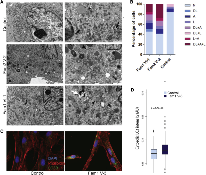
Sphingomyelinases generate ceramide from sphingomyelin as a second messenger in intracellular signaling pathways involved in cell proliferation, differentiation, or apoptosis. Children from 12 unrelated families presented with microcephaly, simplified gyral pattern of the cortex, hypomyelination, cerebellar hypoplasia, congenital arthrogryposis, and early fetal/postnatal demise. Genomic analysis revealed bi-allelic loss-of-function variants in SMPD4, coding for the neutral sphingomyelinase-3 (nSMase-3/SMPD4). Overexpression of human Myc-tagged SMPD4 showed localization both to the outer nuclear envelope and the ER and additionally revealed interactions with several nuclear pore complex proteins by proteomics analysis. Fibroblasts from affected individuals showed ER cisternae abnormalities, suspected for increased autophagy, and were more susceptible to apoptosis under stress conditions, while treatment with siSMPD4 caused delayed cell cycle progression. Our data show that SMPD4 links homeostasis of membrane sphingolipids to cell fate by regulating the cross-talk between the ER and the outer nuclear envelope, while its loss reveals a pathogenic mechanism in microcephaly.

Keywords: NET13; SMPD4; arthrogryposis; microcephaly; neutral-sphingomyelinase.


Conflict of interest statement

The authors declare no competing interests.

Figures

Figure 1
Figure 1
Clinical Phenotype of Individuals with SMPD4 Variants (A) Pedigrees of the 12 families (affected individuals are black symbols). From 21 individuals we obtained both clinical data as well as confirmation of variants in SMPD4. Subjects II-2/9 Fam 3, III-1/2/3/4 Fam 6, VI-1/4 Fam 8, and II-1/2/3 Fam 10 presented with similar clinical features but no extensive clinical reports are available. For 2 of them (Fam 3 II-9 and Fam 8 VI-1), variants in SMPD4 were confirmed by sequencing. (B) Schematic overview of SMPD4 variants (GSDS 2.0). Red arrowheads, truncating variants; purple, intronic splice variants; blue, missense variants. (C) Upper panel: facial features of subject II-1 from family 3 (left), and older IV-1 (middle) and younger IV-2 (right) affected siblings of family 9. Lower panel: facial features of subject III-2 (left) and III-1 (middle) of family 11. Contractions of the distal joints of the upper limbs of subject III-1 (upper) and III-2 (lower) of family 11. (D) Cerebral cortex during gestation. Upper left panel: normal cerebral cortex at the 20th week (hematoxylin and eosin staining, magnification 4×). Upper right panel: cerebral cortex in SMPD4 fetal brain from individual II-2 (family 7) at 22w gestation (cresyl violet staining, magnification 4×). Lower left panel: Normal molecular zone at the 20th week (hematoxylin and eosin staining, magnification 10×). Lower right panel: Molecular zone in SMPD4 case (cresyl violet staining, magnification 10×). Clusters of cells forming dense groups bordering on pial surface and waves of over- migrating neurons (marked with arrows) that disrupt the marginal zone. Lack of subpial granular layer. Marginal zone (MZ), cortical plate (CP), subplate (SP), subpial granular layer (SGL).
Figure 2
Figure 2
Transcriptome Analysis and Quantification of SMPD4 mRNA Expression and Protein Levels (A and B) Graphic illustration (adapted from IGV Sashimi plot) (A) and table of the percentage (B) of SMPD4 transcripts in RNA-seq data of total RNA of individuals Fam 1 V-3 and VI-1 and 4 control individuals. RNA-seq reads are normalized to counts/billion, percentages are calculated for each transcript compared to the total SMPD4 (wild-type and alternative) transcript counts (GRCh38). (C) Allen human brain atlas: expression of nSMases during pre- and postnatal development. Higher expression of SMPD4 in the first 8 weeks post conception (pcw) compared to other nSMases (see Allen Brain Atlas in Web Resources). (D) SMPD4 mRNA expression in fibroblasts from two probands (family 1, VI-1 and family 2, V-2). Unpaired t test with Welch’s correction, two-tailed, n = 2 experiments, 4 independent control cell lines were used. (E) Western blot of SMPD4 in fibroblast homogenate from three individuals with SMPD4 variant (Family 1, VI-1, family 1, V-3 and family 2, V-2) versus two control subjects (C1, C2).
Figure 3
Figure 3
Brain Imaging and Prenatal Ultrasound of Affected Individuals (A–C) Family 1, V-1 at term, T1 weighted images (A, B) and T2 weighted image (C) showing microcephaly (A), simplified gyral pattern (B, C), and insufficient myelination (C). (D–F) T1 images of family 1, VI-4 at term, showing small vermis and simplified gyration. (G–I) T2 weighted images, follow-up MRI of the same subject of family 1, VI-1 at 3 years, few h post-mortem, showing simplified gyration, brain stem hypoplasia and cerebral global hypomyelination. (J–L) Family 2, V-2 (E-A) at 5 years, showing simplified gyral pattern, mild brain stem hypoplasia, thin corpus callosum and global hypomyelination in (L). Note the right parietal positional plagiocephaly. (M–O) Family 3, II-1 (LR00-144) at 3 weeks, showing simplified gyration and delayed myelination on T2 weighted images (N and O). (P–R) Family 9, subject II-2 at the age of 5 years, showing simplified gyral pattern and delayed myelination on T2 weighted images (in R). (S–V) Family 10, subject II-4, prenatal ultrasound at 37 GW. S, clenched hand; T, talipes; U, head circumference corresponding to GW 31+5; V, abdominal circumference corresponding to GW 31+5 (symmetrical IUGR).
Figure 4
Figure 4
Electronmicroscopy of SMPD4-Mutated Fibroblasts and LC3B-II Immunofluorescence Staining (A) Electron microscopy on fibroblasts from individuals with SMPD4 variants and a control subject. Normal granular content of RER is indicated by black arrows in control fibroblasts. Cellular abnormalities in SMPD4-deficient cells include dilated RER cisternae (black arrowheads), with floccular material (white asterisks), increase of lysosomes (ly), and late autophagic vacuoles (white arrow). (B) Quantification of electron microscopy findings; n, normal morphology; dl, dilated ER; A, increased number of autophagic vacuoles; L, increased number of lysomes (n = 153 cells, p < 0.0001 tested by the Fisher and the χ2). (C) Immunofluorescence staining shows increased LC3B-II signal in SMPD4-deficient cells. Phalloidin antibodies mark the actin cytoskeleton, DAPI staining the nucleus. (D) Quantification of LC3B-II cytoplasmic fluorescence signals, one-sided Mann-Whitney U test.
Figure 5
Figure 5
Cell Cycle Analysis and Subcellular Localization of SMPD4 (A) Immunofluorescence stainings of overexpressed Myc-tagged SMPD4 in HEK293T cells. The upper pannel shows localization of Myc-SMPD4 and the ER marker calnexin. The lower panel shows partial colocalization with the nuclear pore marker mab-414. Scale bar represents 5 μm. Partial zoom shows 300% enlargement. (B) Network analysis by IPA (ingenuity platform analysis) of SMPD4 interactors. The proteins found by mass spectometry are depicted in blue. White nodes are connecting proteins not found by mass spectometry. SMPD4 is shown in orange. Solid lines indicate direct interactions, interrupted lines indicate indirect interactions. Network 1 (left hub) shows proteins of the endoplasmic reticulum, mainly involved in post-translational modifications (p = 10e−62). The right network hub shows multiple components of the nuclear pore complex (p = 10e−56). p values are calculated from the network score computed by IPA (Score = -log(Fisher’s extract p value)). (C and D) Flow cytometry of control fibroblasts after downregulation of SMPD4 expression by siRNA (n = 3 experiments, unpaired t test, two tailed) (C) and fibroblasts from control subjects and three individuals with SMPD4 variants (D). G1, S, and G2/M indicate cell cycle phases. The fifth upper graph indicates the level of SMPD4 downregulation achieved. (E) Susceptibility of cultured fibroblasts from individuals with SMPD4 variants to apoptosis when treated with 0.1 mM hydrogen peroxide in comparison to control cell lines, using the FAM-FLICA Caspase-3/7 activity kits. All three available fibroblasts cell lines from SMPD4-affected individuals and two control cell lines were used (n = 3 experiments, p = 0.015, t = 2.60, df = 35, unpaired t test with Welch’s correction, two tailed). Fibroblasts from one individual with Wollcot Rallison syndrome (WRS) were used as a positive control (unpaired t test with Welch’s correction, two tailed, p = 0.0013, t = 5.65, df = 22). On the y axis: percentage of fluorescent cells.

References

    1. Breslow D.K. Sphingolipid homeostasis in the endoplasmic reticulum and beyond. Cold Spring Harb. Perspect. Biol. 2013;5:a013326. - PMC - PubMed
    1. Olsen A.S.B., Færgeman N.J. Sphingolipids: membrane microdomains in brain development, function and neurological diseases. Open Biol. 2017;7:7. - PMC - PubMed
    1. Bienias K., Fiedorowicz A., Sadowska A., Prokopiuk S., Car H. Regulation of sphingomyelin metabolism. Pharmacol. Rep. 2016;68:570–581. - PubMed
    1. Corcoran C.A., He Q., Ponnusamy S., Ogretmen B., Huang Y., Sheikh M.S. Neutral sphingomyelinase-3 is a DNA damage and nongenotoxic stress-regulated gene that is deregulated in human malignancies. Mol. Cancer Res. 2008;6:795–807. - PMC - PubMed
    1. Krut O., Wiegmann K., Kashkar H., Yazdanpanah B., Krönke M. Novel tumor necrosis factor-responsive mammalian neutral sphingomyelinase-3 is a C-tail-anchored protein. J. Biol. Chem. 2006;281:13784–13793. - PubMed

Publication types

Substances