. 2020 Jan:106:102331.
doi: 10.1016/j.jaut.2019.102331. Epub 2019 Sep 5.

Interactions among glomerulus infiltrating macrophages and intrinsic cells via cytokines in chronic lupus glomerulonephritis

Affiliations

Interactions among glomerulus infiltrating macrophages and intrinsic cells via cytokines in chronic lupus glomerulonephritis

Sun-Sang J Sung et al. J Autoimmun. 2020 Jan.

Abstract

Inflammation plays a key role in the pathogenesis of lupus nephritis (LN) and inflammatory cytokines within the glomeruli are critical in this process. However, little information is available for the identities of the cell types that are primarily responsible for the production and function of the various cytokines. We have devised a novel method to visualize cytokine signals in the kidney by confocal microscopy and found that cytokine production within the glomerulus is cell type-specific and under translational control. In the lupus-prone NZM2328 mice with chronic glomerulonephritis, IL-6, IL-1β, and TNF-α in the glomerulus were produced predominantly by mesangial cells, podocytes, and glomerulus-infiltrating blood-derived macrophages, respectively. Microarray and RNASeq analyses showed that these cells expressed the receptors for these cytokines. Together the 3 cell types form a cytokine circuit in amplifying cytokine responses in LN. The intrinsic cells and infiltrating macrophages also produced other cytokines including M-CSF, SCF, and IL-34 that constituted within the enclosed glomerular space the soluble effector milieu which may mediate cellular damage and proliferation, and cytokine transcriptional and translation regulation. IL-10 and IL-1β were translationally regulated in the glomeruli in the intact kidney in a cell type-specific manner. The production of these 2 cytokines by infiltrating macrophages was undetectable in a visualization system for in situ protein accumulation despite high mRNA expression levels. However, these macrophages in isolated glomeruli which are released from Bowman's capsules produced large amounts of IL-10 and IL-1β. These data reveal the complexity of cytokine regulation, production, and function in the glomerulus and provide a model in which cytokine blocking may be beneficial in LN treatment.

Keywords: Cytokines; Lupus nephritis; Macrophages; Mesangial cells; Podocytes; Translational regulation.


Figures

Fig. 1.
Fig. 1.
Cytokine mRNA expression by glomerulus-infiltrating macrophages. Glomerular CD11b+ infiltrating macrophages in 3 Mo old NZM mice with trace proteinuria by dipstick (light gray) or 7 Mo – 12 Mo old NZM mice with 3+ - 4+ proteinuria (dark gray) were purified by cell sorting of single cell suspensions of magnetic bead-isolated glomeruli as described in Materials and Methods. In each sort, glomerular cells from 3-4 mice were pooled. Total RNA were extracted and subjected to reverse transciptase-real time PCR analysis. The results are pooled from 2 – 4 experiments. *, p < 0.05.
Fig. 2.
Fig. 2.
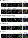
Glomerulus-infiltrating macrophages in NZM mice with cGN produce TNF-α but not IL-6 and IL-1β cytokine protein. Mice with 3+ to 4+ proteinuria by dipstick were perfused with 20 ml of medium with PMA, ionomycin, brefeldin A, and monensin and kidney slices were incubated in the same medium with 10% hiFCS for 6 h. Tissues slices were fixed in PLP, passed through a sucrose gradient, and embedded in OCT. Tissue sections (5 μm) were stained simultaneously with 3 – 4 Ab directly conjugated with fluorochromes of excitation wavelengths of 421, 488, 555, and 647 nm. Photomicrographs were collected on a Zeiss LSM-700 confocal microscope assembly and analyzed by the program ZEN. At least 5 mice were analyzed by the same Ab combinations and >15 glomeruli were recorded for each determination. (A) Arrows show selected CD11b+ macrophages producing TNF-α. (B) Arrowheads show that TNF-α staining (panels a, e) is not associated with mesangial cell (panel b, e, f) or endothelial cell (panels c, e, f) staining. (C) CD11b+ macrophages did not produce IL-6 (arrows show macrophages, panels a, b, e, f). (D) Arrowheads show CD11b+ macrophages with no IL-1β production (panels b, e, f). In panels f, glomeruli were shown in circles. Bars in f equals 10 μm.
Fig. 3.
Fig. 3.
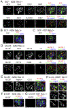
IL-6 production by mesangial cells in NZM mice with cGN. Kidney slices from NZM or R27 mice with and without severe proteinuria (3+ dipstick) were stimulated in the presence of secretion blockers as described in Fig. 2. Sections were stained with the indicated fluorochrome-conjugated Ab. Photomicrographs were captured by confocal microcopy and analyzed by ZEN software. (A) IL-6 staining colocalized with mesangial cell staining (arrows, panels a, b, e, yellow in e) but not with endothelial cells (arrows, panels c, e) and CD45+ leukocytes (arrowheads, panels d, f). (B) IL-6 (arrowheads, panels a, e, f) is not produced by podocytes, CD11b macrophages, and I-A+ macrophages/DC (arrowheads, panels b-f). IL-6 is not detected in the glomeruli of young NZM mice (C) and R27 mice of all ages (E). In old NZM mice without cGN, few mesangial cells (arrows in D, panels a-c) produced IL-6. Bars are 10 μm.
Fig. 4.
Fig. 4.
Podocytes produced IL-1β in kidneys of NZM mice with severe proteinuria and cGN. Tissue slices were processed and stained as described in Fig. 2. (A) IL-1β staining is associated exclusively with podocytes (panels a, b, d, e; colocalization, yellow in d and e) and not with endothelial cells (panels a, c, d; blue vs. yellow in d and e) and I-A+ cells (panel e, blue vs. yellow). No IL-1β staining (panel f, i, j, red) was associated with mesangial cells (panels g, and i, and j, green). Endothelial cells and I-A+ cells are shown in blue in panels i, and j respectively. In A, panels a-e show one glomerulus and f-j show a second glomerulus. (B) Little IL-1β was detected in kidneys of NZM mice with severe proteinuria and cGN in the absence of stimulation and secretion blocking (IL-1β, arrows). (C) Small amounts of IL-1β was found associated with podocytes in young NZM mice with no proteinuria (IL-1β, panels a, d, e) and in (D) R27 mice at age 6 Mo to 12 Mo with little proteinuria (IL-1β, panels a, d, and e). Bars in panels e equals 10 μm.
Fig. 5.
Fig. 5.
Cytokine and cytokine receptor mRNA expression by mesangial cells, renal myeloid cells, and podocytes. (A, B) Mesangial cells, and interstitial macrophages and dendritic cells from NZM mice with 3+ - 4+ proteinuria were purified by FACS sorting as described in Materials and Methods and used as the RNA source for microarray analysis. Heat maps of growth factors and growth factor receptors are shown in (A) and (B) respectively. Three independent sorts with pools of 4-5 mice each was used. (C, D) Heat map of podocyte cytokine and cytokine receptor mRNA expression by RNASeq. The data was obtained from GEO dataset GSE64063 [11].
Fig. 6.
Fig. 6.
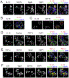
Mesangial cells produce SCF and M-CSF and podocytes produce IL-34 in NZM mice with cGN. Kidney tissues were prepared as described in Fig. 2 and stained with the indicated fluorochrome-conjugated Ab. (A) SCF staining (arrows, panel a, e, f) colocalizes with mesangial cell staining (panels b, e, f yellow in e, f) but not with Mac2+ macrophages (panels h, k, l) or proximal or distal tubular cells (panel i – l). Panels a-f is from 1 glomerulus and g-l is from a second glomerulus. Glomerular cells in young NZM mice (B) and 6-12 Mo old adult R27 mice (C) with no proteinuria did not produce SCF (red). (D) M-CSF (arrows, panels a-c, e, f, red to yellow in e, f due to cytoplasmic vs. membrane staining) was produced by mesangial cells (arrows, panel b; green in panels e, f) but not by CD11b+ macrophages (panel c; blue, panel e) or I-A+ macrophage/DC (panel d, blue, panel f). F4/80+ macrophages also did not produce M-CSF (panels i, k). (E) Podocytes (panels b, d) and glomerular endothelial cells (panels c, d) in NZM mice with cGN did not produce M-CSF (panels a, d). Young NZM mice (F) and 6-12 Mo old R27 mice (G) without cGN did not produce M-CSF. (H) IL-34 production by podocytes in NZM mice with cGN. Arrows shows colocalization (yellow) of IL-34 (red in a, b) with podocytes (green in a, b). The individual staining of IL-34, nephrin, Itgα8, and CD31 are shown in c, d, e, and f respectively. Glomeruli are outlined in white. Bars equal 10 μm.
Fig. 7.
Fig. 7.
High levels of IL-10, IL-1β and TNF-α production by CD11b+ macrophages with little IL-6 production in isolated glomeruli. Glomeruli were isolated from NZM mice with cGN by magnetic beads prior to stimulation with PMA and ionomycin, plus secretion blocking with brefeldin A and monensin. The glomeruli were fixed in 2% PLP, equilibrated in sucrose gradient, and frozen in OCT. Sections were stained with fluorochrome-conjugated Ab against cytokines and glomerular intrinsic cell and macrophage markers. (A) In isolated glomeruli, IL-10 was produced by CD11b+ macrophages but not mesangial cells (arrows, IL-10 and macrophages, panels a, b, e, f; yellow for colocalization in e, f). On the other hand, IL-10 was not detected in glomerular macrophages and intrinsic cells when cells were stimulated in tissue slices (B; arrows indicate CD11b+ macrophages). (C) Similarly, IL-1β (panels a, c, red in c) was not produced by CD11b+ macrophages in tissues (arrows, macrophages; panel c, green) whereas large amounts of IL-1β (panels a, c, red in c) was produced by podocytes. In contrast, in isolated decapsulated glomeruli (D), macrophages produced large amounts of IL-1β (arrows, IL-1β and macrophages in panels a, c, e; magenta for IL-1β and CD11b colocalization in e). Lower levels of podocyte IL-1β production was detected (arrowheads, panels a, b, e, f; yellow for podocyte and IL-1β colocalizaiton in e, f). (E) TNF-α was produced exclusively by CD11b+ macrophages (arrows, TNF-α and macrophages, panels a, b, e, f; yellow for CD11b+ TNF-α+ cells in e, f). (F) IL-6 was detectable only in Itgα8+ mesangial cells (arrows, IL-6 and mesangial cells, panels a, b, e, f; yellow for Itgα8+IL-6+ cells). Glomeruli are enclosed in white outlines and bars equal 10 μm. These experiments are repeated 3 times with 2-3 NZM mice with cGN in each experiment.

References

    1. Fu SM, Sung S.-s. J, Wang H, Gaskin F, Pathogenesis of Lupus Nephritis, in: Wallace D, Hahn B, (Eds.), Dubois’ Lupus Erythematosus and Related Syndromes, 9th Edition, Elsevier, New York, N.Y., 2019, pp. 269–293.
    1. Tsokos GC, Systemic lupus erythematosus, N. Engl. J. Med 365 (2011) 2110–2121. 10.1056/NEJMra1100359. - DOI - PubMed
    1. Greka A, Mundel P, Cell biology and pathology of podocytes, Annu. Rev. Physiol 74 (2012) 299–323. 10.1146/annurev-physiol-020911-153238. - DOI - PMC - PubMed
    1. Brunskill EW, Aronow BJ, Georgas K, Rumballe B, Valerius MT, Aronow J et al., Atlas of gene expression in the developing kidney at microanatomic resolution, Developmental cell 15 (2008) 781–791. 10.1016/j.devcel.2008.09.007. - DOI - PMC - PubMed
    1. Naito Y, Uchiyama K, Mizushima K, Kuroda M, Akagiri S, Takagi T et al., Microarray profiling of gene expression patterns in glomerular cells of astaxanthin-treated diabetic mice: a nutrigenomic approach, Int. J. Mol. Med 18 (2006) 685–695 - PubMed

Publication types