. 2019 Nov;23(11):7819-7829.
doi: 10.1111/jcmm.14661. Epub 2019 Sep 9.

Schistosoma japonicum peptide SJMHE1 suppresses airway inflammation of allergic asthma in mice

Affiliations

Schistosoma japonicum peptide SJMHE1 suppresses airway inflammation of allergic asthma in mice

Wenzhe Zhang et al. J Cell Mol Med. 2019 Nov.

Abstract

Helminths and their products can shape immune responses by modulating immune cells, which are dysfunctional in inflammatory diseases such as asthma. We previously identified SJMHE1, a small molecule peptide from the HSP60 protein of Schistosoma japonicum. SJMHE1 can inhibit delayed-type hypersensitivity and collagen-induced arthritis in mice. In the present study, we evaluated this peptide's potential intervention effect and mechanism on ovalbumin-induced asthma in mice. SJMHE1 treatment suppressed airway inflammation in allergic mice, decreased the infiltrating inflammatory cells in the lungs and bronchoalveolar lavage fluid, modulated the production of pro-inflammatory and anti-inflammatory cytokines in the splenocytes and lungs of allergic mice, reduced the percentage of Th2 cells and increased the proportion of Th1 and regulatory T cells (Tregs). At the same time, Foxp3 and T-bet expression increased, and GATA3 and RORγt decreased in the lungs of allergic mice. We proved that SJMHE1 can interrupt the development of asthma by diminishing airway inflammation in mice. The down-regulation of Th2 response and the up-regulation of Th1 and Tregs response may contribute to the protection induced by SJMHE1 in allergic mice. SJMHE1 can serve as a novel therapy for asthma and other allergic or inflammatory diseases.

Keywords: SJMHE1; Schistosoma japonicum peptide; airway inflammation; allergic asthma; suppress.


Conflict of interest statement

The authors declare that they have no competing interests. The funding agencies played no role in the design or implementation of the study, analysis or interpretation of the data, or the preparation and submission of the manuscript.

Figures

Figure 1
Figure 1
SJMHE1 treatment inhibits the development of airway inflammation in allergic mice. A, Experimental scheme. BALB/c mice were sensitized with OVA on days 0, 7 and 14 and challenged with OVA daily from days 21 to 27. Mice were injected with SJMHE1 emulsified in IFA on days 0, 14 and day 28. The mice were killed on day 35. B, Histological analysis of lung section from mice by H&E staining. 20× magnification. Images are representative of four independent experiments (n = 6 mice per group). C, Inflammatory process was scored following Underwood's standards, with 15 indicating the most severe pathological changes for each mouse. D, Total cells in BALF. E, Eosinophil number in BALF. The quantity of eosinophil in BALF cells was determined following Wright and Giemsa staining. F, OVA‐specific IgE antibody levels in sera of mice. Results are presented as mean ± SEM (n = 12) from two‐independent experiments. *** P < .001
Figure 2
Figure 2
SJMHE1 treatment modulates the expression of cytokines from splenocytes of allergic mice. On day 35, the mice were killed, and the splenocytes from each mouse were tested for mRNA expression of IFN‐γ (A), IL‐4 (B), IL‐17 (C), IL‐10 (D), TGF‐β (E) and IL‐35 (F) by qRT‐PCR. Results are presented as mean ± SEM of 12 mice from two independent experiments performed in triplicate wells. * P < .05, *** P < .001
Figure 3
Figure 3
SJMHE1 treatment modulates the expression of cytokines from lungs of allergic mice. On day 35, the mice were killed, and the lungs from each mouse were tested for mRNA expression of IFN‐γ (A), IL‐4 (B), IL‐5 (C), IL‐17 (D), IL‐10 (E), TGF‐β (F) and IL‐35 (G) by qRT‐PCR. Results are presented as mean ± SEM of 12 mice from two independent experiments performed in triplicate wells. * P < .05, ** P < .01, *** P < .001
Figure 4
Figure 4
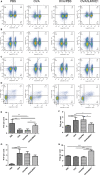
SJMHE1 treatment modulates Th1/Th2/Th17/Treg response in splenocytes of allergic mice. On day 35, the mice were killed, and the splenocytes from each mouse were tested for Th1/Th2/Th17/Treg subsets by flow cytometry. A, CD4+IFN‐γ+ Th1 cells, B, CD4+IL4+ Th2 cells, C, CD4+IL17+ Th17 cells and D, CD4+CD25+Foxp3+ Tregs in each group are shown. Data are representative of the experiments. E, The percentage of CD4+IFN‐γ+ Th1 cells, F, the percentage of CD4+IL4+ Th2 cells, G, the percentage of CD4+IL17+ Th17 cells and H, the percentage of CD4+CD25+Foxp3+ Tregs in each group are shown. Results are presented as mean ± SEM of 12 mice from two independent experiments. * P < .05, ** P < .01, *** P < .001
Figure 5
Figure 5
SJMHE1 treatment increases the expression of Foxp3 in splenocytes and lungs of allergic mice. On day 35, the mice were killed, and the splenocytes and lungs from each mouse were tested for Foxp3 expression. A, Foxp3 expression in lungs by IHC staining. Images are representative of two independent experiments (n = 6 mice per group). B, Quantification of Foxp3‐positive areas in each group using Image‐Pro Plus software. Data are presented as mean ± SEM of 12 mice from two independent experiments. C, The expression of Foxp3 mRNA in splenocytes and lungs D, from each group by qRT‐PCR. Data are presented as mean ± SEM of 12 mice from two independent experiments. ** P < .01, *** P < .001
Figure 6
Figure 6
SJMHE1 treatment up‐regulates the expression of T‐bet and down‐regulates the expression of GATA3 and RORγt in lungs of allergic mice. On day 35, the mice were killed, and the lungs from each mouse were tested for T‐bet, GATA3 and RORγt expression by Western blotting analysis. A representative blot from each group is shown

Similar articles

Cited by

References

    1. Lemanske RF Jr, Busse WW. Asthma: clinical expression and molecular mechanisms. J Allergy Clin Immunol. 2010;125:S95‐S102. - PMC - PubMed
    1. Muehling LM, Lawrence MG, Woodfolk JA. Pathogenic CD4(+) T cells in patients with asthma. J Allergy Clin Immunol. 2017;140:1523‐1540. - PMC - PubMed
    1. Shi YH, Shi GC, Wan HY, et al. Coexistence of Th1/Th2 and Th17/Treg imbalances in patients with allergic asthma. Chin Med J. 2011;124:1951‐1956. - PubMed
    1. Gilchrist FJ, Ahmad AN, Batchelor HK, Marriott JF, Lenney W. A review of prednisolone prescribing for children with acute asthma in the UK. J Asthma. 2016;53:563‐566. - PubMed
    1. Choby GW, Lee S. Pharmacotherapy for the treatment of asthma: current treatment options and future directions. Int Forum Allergy Rhinol. 2015;5(Suppl 1):S35‐40. - PubMed

Publication types

MeSH terms