Review
. 2019 Aug 21;25(31):4383-4404.
doi: 10.3748/wjg.v25.i31.4383.

Systemic inflammation in colorectal cancer: Underlying factors, effects, and prognostic significance

Affiliations
Review

Systemic inflammation in colorectal cancer: Underlying factors, effects, and prognostic significance

Anne E Tuomisto et al. World J Gastroenterol. .

Abstract

Systemic inflammation is a marker of poor prognosis preoperatively present in around 20%-40% of colorectal cancer patients. The hallmarks of systemic inflammation include an increased production of proinflammatory cytokines and acute phase proteins that enter the circulation. While the low-level systemic inflammation is often clinically silent, its consequences are many and may ultimately lead to chronic cancer-associated wasting, cachexia. In this review, we discuss the pathogenesis of cancer-related systemic inflammation, explore the role of systemic inflammation in promoting cancer growth, escaping antitumor defense, and shifting metabolic pathways, and how these changes are related to less favorable outcome.

Keywords: C-reactive protein; Cachexia; Chemokine; Colorectal cancer; Cytokine; Glasgow prognostic score; Inflammation; Metastasis; Prognosis.


Conflict of interest statement

Conflict-of-interest statement: The authors declare that there is no conflict of interest.

Figures

Figure 1
Figure 1
Phenotypic spectrum of macrophages. The illustration portrays the heterogeneity of immune cell main categories in producing inflammatory mediators and growth factors based on their activation state. The image illustrates the spectrum model of macrophage polarization based on the M1-M2 paradigm. Macrophage polarization describes the type of macrophage activation at a given point in space and time[155]. The polarization can be viewed as a continuum, with M1 (pro-inflammatory) and M2 (anti-inflammatory) as the extremes. The M1 and M2 designations are based on in vitro stimulation with either interferon gamma (M1) or interleukin 4 (M2) without environmental influence[54]; in vivo, stimulation of macrophages with multiple cytokines may result in mixed phenotypes. The image shows examples of transcription factors, cell surface molecules and inflammatory mediators commonly associated with M1 and M2 polarization states. Similarly to macrophages, different activation states have been associated with other immune cell types such as neutrophils[57,58], B cells[56], and plasma cells[156]. CCL: C-C motif chemokine ligand; CD80: CD80 molecule; CD86: CD86 molecule; CXCL: C-X-C motif chemokine ligand; GATA3: GATA binding protein 3; IL: Interleukin; IRF: Interferon regulatory factor; MHC II: Major histocompatibility complex, type II; MMP1: Matrix metallopeptidase 1; MRC1; Mannose receptor C-type 1; pSTAT1: Phosphorylated signal transducer and activator of transcription 1; STAB1: Stabilin 1; TGFB1: Transforming growth factor beta 1; TNF: Tumor necrosis factor; VEGFA: Vascular endothelial growth factor A.
Figure 2
Figure 2
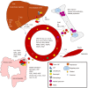
Overview of the effects of systemic inflammation in colorectal cancer. The illustration portrays some of the molecules and phenomena considered important in the pathogenesis of colorectal cancer associated systemic inflammation. Some markers showing increased circulating concentrations in colorectal cancer patients are listed in the center. ALB: Albumin; CCL: C-C motif chemokine ligand; CXCL: C-X-C motif chemokine ligand; CRP: C-reactive protein; CSF: Colony stimulating factor; FGF2: Fibroblast growth factor 2; Gln: Glutamine; HP: Haptoglobin; IL: Interleukin; MMP: Matrix metallopeptidase; OPN: Osteopontin; PDGF: Platelet derived growth factor; SAA1: Serum amyloid A1; THPO: Thrombopoietin; TIMP1: TIMP metallopeptidase inhibitor 1; VEGFA: Vascular endothelial growth factor A; VEGFC: Vascular endothelial growth factor C; vWF: von Willebrand factor
Figure 3
Figure 3
Overview of the potential pathways involved in the suppression of anti-tumor immunity by myeloid derived suppressor cells. ARG1: Arginase 1; IDO1: Indoleamine 2,3-dioxygenase 1; IL10: Interleukin 10; MDSCs: Myeloid derived suppressor cells; PD1: Programmed cell death protein 1; PDL1: Programmed death ligand 1; TGFB1: Transforming growth factor beta 1.

Similar articles

Cited by

References

    1. Hanahan D, Weinberg RA. Hallmarks of cancer: the next generation. Cell. 2011;144:646–674. - PubMed
    1. McAllister SS, Weinberg RA. Tumor-host interactions: a far-reaching relationship. J Clin Oncol. 2010;28:4022–4028. - PubMed
    1. Schreiber RD, Old LJ, Smyth MJ. Cancer immunoediting: integrating immunity's roles in cancer suppression and promotion. Science. 2011;331:1565–1570. - PubMed
    1. Balkwill FR, Capasso M, Hagemann T. The tumor microenvironment at a glance. J Cell Sci. 2012;125:5591–5596. - PubMed
    1. McAllister SS, Weinberg RA. The tumour-induced systemic environment as a critical regulator of cancer progression and metastasis. Nat Cell Biol. 2014;16:717–727. - PMC - PubMed

MeSH terms