. 2019 Aug 9:12:6297-6307.
doi: 10.2147/OTT.S214689. eCollection 2019.

LncRNA-MALAT1 regulates proliferation and apoptosis of ovarian cancer cells by targeting miR-503-5p

Affiliations

LncRNA-MALAT1 regulates proliferation and apoptosis of ovarian cancer cells by targeting miR-503-5p

Qian Sun et al. Onco Targets Ther. .

Abstract

Objective: Ovarian cancer (OC) is a common female disease with a poor prognosis. But the possible mechanism of OC tumor progression remains an active area of research. This study is intended to explore the effect of metastasis-associated lung adenocarcinoma transcript 1 (MALAT1) on proliferation and apoptosis of OC and its mechanism.

Materials and methods: MALAT1 and miR-503-5p expressions in human OC cell lines and normal human ovarian epithelial (HOSE) cell line were measured using qRT-PCR. OC cell line SKOV3 is divided into 4 groups: pcDNA3.1 group, pcDNA3.1-MALAT1 group, si-NC group, and si-MALAT1 group. MTT assay and 5-ethynyl-2'-deoxyuridine (EdU) assay were applied for the detection of cell proliferation. Relationship of MALAT1 with miR-503-5p was verified using luciferase assay and RNA pull-down. The luciferase activity in cells was normalized to RNA concentrations determined by Bradford assays.

Results: MALAT1 expression in OC cells was elevated compared with HOSE cells. MTT assay and EdU assay supported that si-MALAT1 could inhibit cell proliferation in OC cells. Treatment of si-MALAT1 results in increased cell apoptosis rate in both SKOV3 cells and OVCAR3 cells. The expression of lncRNA-MALAT1 was negatively associated with the expression of miR-503-5p in OC cells, while luciferase assay and RNA pull-down together supported the direct binding of MALAT1 with miR-503-5p. Knockdown of MALAT1 was able to inhibit the activation of JAK2/STAT3 signal pathway, and MALAT1 overexpression was accompanied by activation of these factors.

Conclusion: lncRNA-MALAT1 can negatively target miR-503-5p expression to further promote proliferation and depress apoptosis of OC cells through the JAK2-STAT3 pathway.

Keywords: JAK2/STAT3; cell apoptosis; cell proliferation; lncRNA-MALAT1; miR-503-5p; ovarian cancer.


Conflict of interest statement

The authors report no conflicts of interest in this work.

Figures

Figure 1
Figure 1
MALAT1 was upregulated in ovarian cancer cells relative to normal ovarian epithelial cells, and RNAi downregulated MALAT1 expression. Notes: (A) The relative expression level of MALAT1 was detected by reverse transcription-quantitative polymerase chain reaction analysis in four ovarian cancer cell lines (CaOV3, SKOV3, OVCAR3, and OV90) and a normal ovarian epithelial cell line (HOSE). **P<0.01, ***P<0.001 vs HOSE. (B) The fold changes of MALAT1 expression in SKOV3 and OVSAR3 cells were analyzed 48 hrs after treating with pcDNA3.1, pcDNA3.1-MALAT1, si-MALAT1, or si-NC. Data are presented as the mean ± standard error of the mean. Experiments were performed in triplicate. **P<0.01 vs si-NC transfections. Abbreviations: MALAT1, metastasis-associated lung adenocarcinoma transcript 1; si-MALAT1, MALAT1 small interfering RNA; si-NC, noncoding small interfering RNA.
Figure 2
Figure 2
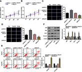
MALAT1 promoted cell proliferation and inhibited cell apoptosis in SKOV3 and OVCAR3 cells. Notes: The cell growths of SKOV3 (A) and OVCAR3 (B) cells were monitored using MTT assays in response to si-MALAT1 or pcDNA3.1-MALAT1 induced MALAT1 knockdown or overexpression, respectively. Effects of MALAT1 knockdown or overexpression on cell proliferations of SKOV3 (C) and OVCAR3 (D) were further determined by Edu staining (200×). (E) Apoptosis of SKOV3 and OVCAR3 cells transfected with si-MALAT1, si-NC, pcDNA3.1-MALAT1, or pcDNA3.1 was analyzed by flow cytometry. The percentage displayed on the histograms indicates apoptotic rate. Comparison of apoptotic rates of cells transfected with si-MALAT1, si-NC, pcDNA3.1-MALAT1, or pcDNA3.1. (F) Proteins were detected by Western blot. β-Actin was used as internal control to ensure equal loadings. Graphs are represented as the mean density of p53, Bax, Bcl-2, and Survivin bands normalized against the mean density of β-actin band from three independent experiments (presented as relative density of individual protein). Data are presented as the mean ± standard error of the mean (n=3). *P<0.05, **P<0.01, ***P<0.001  vs si-NC, or pcDNA3.1. Abbreviations: MALAT1, metastasis-associated lung adenocarcinoma transcript 1; si-MALAT1, MALAT1 small interfering RNA; si-NC, noncoding small interfering RNA.
Figure 3
Figure 3
MALAT1 directly targets miR-503-5p. Notes: (A) The expressions of miR-503-5p in SKOV3 and OVCAR3 cells were determined in response to overexpression or knockdown of MALAT1 using reverse transcription-quantitative polymerase chain reaction analysis. (B) MALAT1 expression in response to miR-503-5p overexpression or miR-503-5p inhibition was determined by using real-time PCR. (C) Mimics NC/miR-503-5p mimics or inhibitor NC/miR-503-5p inhibitor was transfected into SKOV3 and OVCAR3 cells to achieve miR-503-5p overexpression or inhibition, verified by using real-time PCR. (D) A WT-MALAT1 luciferase reporter vector (WT-MALAT1) and a MT-MALAT1 luciferase reporter vector (MT-MALAT1) with mutations on miR-503-5p binding sites of the MALAT1 were constructed. (E) The WT-MALAT1/MT-MALAT1 vectors and miR-503-5p NC/miR-503-5p mimics/miR-503-5p inhibitor were co-transfected into SKOV3 and OVCAR3 cells. The luciferase activity of the MALAT1 luciferase reporter vector was determined. (F) Verification of the binding of MALAT1 and miR-503-5p determined by the pull-down assay using MALAT1 probe. (G) Verification of the binding of MALAT1 and miR-503-5p determined by the pull-down assay using miR-503-5p probe. Scram probe was used as negative control. Data are presented as the mean ± standard error of the mean (n=3). *P<0.05, **P<0.01, ***P<0.001  vs NC or blank control. Abbreviations: MALAT1, metastasis-associated lung adenocarcinoma transcript 1; si-MALAT1, MALAT1 small interfering RNA; si-NC, noncoding small interfering RNA; NB, Northern blot.
Figure 4
Figure 4
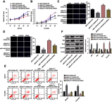
MALAT1 regulated OC cell proliferation and apoptosis through interaction with miR-503-5p. Notes: (A and B) The cell growth was monitored in response to co-processing MALAT1 knockdown and miR-503-5p inhibition using MTT assay in both SKVO3 and OVCAR3 cells. (C and D), Edu staining was applied to observe the co-effect of MALAT1 knockdown and miR-503-5p inhibition on cell proliferation on SKVO3 and OVCAR3 cells (200×). (E) Apoptosis of SKOV3 and OVCAR3 cells co-transfected si-MALAT1/si-NC and miR-503-5p inhibitor/inhibitor NC was analyzed by flow cytometry. The percentage displayed on the histograms indicates apoptotic rate. Comparison of the apoptotic rates of cells co-transfected with si-MALAT1/si-NC and miR-503-5p inhibitor/inhibitor NC. (F) Apoptotic or antiapoptotic proteins were detected by Western blot. β-Actin was used as internal control to ensure equal loadings. Graphs are represented as the mean density of p53, Bax, Bcl-2, and Survivin bands normalized against the mean density of β-actin band from three independent experiments (presented as relative density of individual protein). Data are presented as the mean ± standard error of the mean (n=3). *P<0.05, **P<0.01 vs si-NC or blank control. Abbreviations: MALAT1, metastasis-associated lung adenocarcinoma transcript 1; si-MALAT1, MALAT1 small interfering RNA; si-NC, noncoding small interfering RNA.
Figure 5
Figure 5
MALAT1 activated JAK2/STAT3 pathway. Notes: (A) Total cell lysates were collected and immunoblotted with the indicated antibodies. β-Actin was used as a loading control. Graphs are presented as the mean density of p-JAK2 and p-STAT3 normalized against the mean density of JAK2 or STAT3 from three independent experiments (presented as relative density of phosphoprotein vs total protein). Data are presented as the mean ± standard error of the mean (n=3). *P<0.05, **P<0.01, ***P<0.001 compared to untreated control cells at 0 min. Abbreviations: MALAT1, metastasis-associated lung adenocarcinoma transcript 1; si-MALAT1, MALAT1 small interfering RNA; si-NC, noncoding small interfering RNA.

Similar articles

Cited by

References

    1. Siegel R, Naishadham D, Jemal A. Cancer statistics, 2013. CA Cancer J Clin. 2013;63:11–30. doi:10.3322/caac.21166 - DOI - PubMed
    1. Suh DH, Kim JW, Kim K, Kim HJ, Lee KH. Major clinical research advances in gynecologic cancer in 2012. J Gynecol Oncol. 2013;24:66–82. doi:10.3802/jgo.2013.24.1.66 - DOI - PMC - PubMed
    1. Jelovac D, Armstrong DK. Recent progress in the diagnosis and treatment of ovarian cancer. CA Cancer J Clin. 2011;61:183–203. doi:10.3322/caac.20113 - DOI - PMC - PubMed
    1. Shapira I, Oswald M, Lovecchio J, et al. Circulating biomarkers for detection of ovarian cancer and predicting cancer outcomes. Br J Cancer. 2014;110:976–983. doi:10.1038/bjc.2013.795 - DOI - PMC - PubMed
    1. Lengyel E. Ovarian cancer development and metastasis. Am J Pathol. 2010;177:1053–1064. doi:10.2353/ajpath.2010.100105 - DOI - PMC - PubMed