

Long noncoding RNA HCP5 suppresses skin cutaneous melanoma development by regulating RARRES3 gene expression via sponging miR-12
- PMID: 31496735
- PMCID: PMC6698080
- DOI: 10.2147/OTT.S195796
Long noncoding RNA HCP5 suppresses skin cutaneous melanoma development by regulating RARRES3 gene expression via sponging miR-12
Abstract
Objective: This research aimed to investigate the role and mechanism of long noncoding RNA (lncRNA) HCP5 in skin cutaneous melanoma (SKCM).
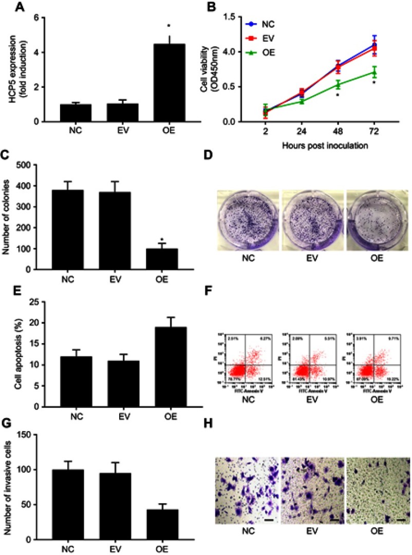
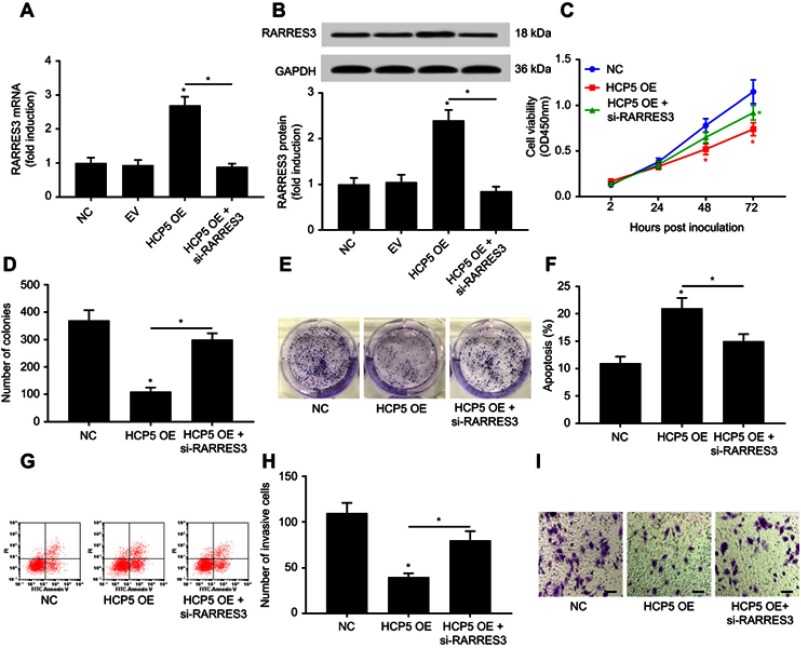
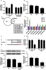
Materials and methods: Survival analysis was performed using The Cancer Genome Atlas (TCGA)-SKCM data and SKCM patients' clinical data. Primary SKCM cells were derived from patients' pathologic tissue specimens. HCP5 overexpression was achieved by lentiviral transduction. Malignancy of SKCM cells was evaluated in vitro by cell proliferation, colony formation, apoptosis and transwell invasion assays. RARRES3 knockdown was achieved by siRNA transfection. DIANA microT-CDS algorithm was used to predict miRNAs that might interact with HCP5 and 3' untranslated region of RARRES3 mRNA. microRNA target luciferase reporter assay and AGO2-RNA immunoprecipitation were used to verify the interaction between HCP5, 3' UTR of RARRES3 mRNA and miR-1286.
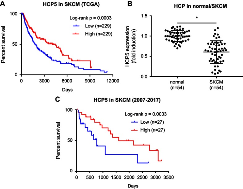
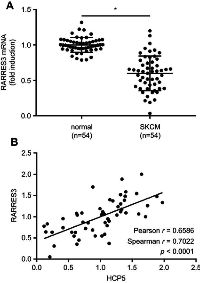
Results: HCP5 level was decreased in SKCM tissue specimens compared to noncancerous counterparts. Low expression of HCP5 was associated with SKCM patients' poor overall survival and disease progression. HCP5 overexpression significantly reduced the malignancy of primary SKCM cells in vitro. RARRES3 was found as a HCP5-co-expressing gene in SKCM cells. HCP5 overexpression significantly increased RARRES3 expression in SKCM cells. RARRES3 knockdown partially abolished the anti-SKCM effect of HCP5 overexpression. MiR-1286 was found interacting with both HCP5 and 3' UTR of RARRES3 mRNA.
Conclusion: HCP5 is a cancer-suppressive lncRNA in SKCM. HCP5 overexpression decreased SKCM cell malignancy in vitro by upregulating RARRES3, possibly via sponging miR-1286.
Keywords: HCP5; RARRES3; long noncoding RNA; miR-1286; skin cutaneous melanoma.
Conflict of interest statement
The authors report no conflicts of interest in this work.
Figures
Similar articles
-
LncRNA HCP5 as a potential therapeutic target and prognostic biomarker for various cancers: a meta‑analysis and bioinformatics analysis.Cancer Cell Int. 2021 Dec 19;21(1):686. doi: 10.1186/s12935-021-02404-x. Cancer Cell Int. 2021. PMID: 34923990 Free PMC article. Review.
-
LncRNA HCP5 Regulates Pancreatic Cancer Progression by miR-140-5p/CDK8 Axis.Cancer Biother Radiopharm. 2020 Nov;35(9):711-719. doi: 10.1089/cbr.2019.3294. Epub 2020 May 14. Cancer Biother Radiopharm. 2020. PMID: 32407143
-
Knockdown of HCP5 exerts tumor-suppressive functions by up-regulating tumor suppressor miR-128-3p in anaplastic thyroid cancer.Biomed Pharmacother. 2019 Aug;116:108966. doi: 10.1016/j.biopha.2019.108966. Epub 2019 May 16. Biomed Pharmacother. 2019. PMID: 31102936
-
Long Noncoding RNA HCP5 Regulates Pancreatic Cancer Gemcitabine (GEM) Resistance By Sponging Hsa-miR-214-3p To Target HDGF.Onco Targets Ther. 2019 Oct 4;12:8207-8216. doi: 10.2147/OTT.S222703. eCollection 2019. Onco Targets Ther. 2019. PMID: 31632071 Free PMC article.
-
Silencing of long non-coding RNA HCP5 inhibits proliferation, invasion, migration, and promotes apoptosis via regulation of miR-299-3p/SMAD5 axis in gastric cancer cells.Bioengineered. 2021 Dec;12(1):225-239. doi: 10.1080/21655979.2020.1863619. Bioengineered. 2021. PMID: 33371778 Free PMC article.
Cited by
-
Exploring Potential Therapeutic Applications of Tazarotene: Gene Regulation Mechanisms and Effects on Melanoma Cell Growth.Curr Issues Mol Biol. 2025 Mar 28;47(4):237. doi: 10.3390/cimb47040237. Curr Issues Mol Biol. 2025. PMID: 40699636 Free PMC article. Review.
-
Using Immune-Related lncRNA Signature for Prognosis and Response to Immunotherapy in Cutaneous Melanoma.Int J Gen Med. 2021 Oct 8;14:6463-6475. doi: 10.2147/IJGM.S335266. eCollection 2021. Int J Gen Med. 2021. PMID: 34675614 Free PMC article.
-
Clinical implications and molecular mechanism of long noncoding RNA LINC00518 and protein-coding genes in skin cutaneous melanoma by genome‑wide investigation.Arch Dermatol Res. 2025 Feb 22;317(1):454. doi: 10.1007/s00403-025-03961-1. Arch Dermatol Res. 2025. PMID: 39987414
-
LncRNA HCP5 as a potential therapeutic target and prognostic biomarker for various cancers: a meta‑analysis and bioinformatics analysis.Cancer Cell Int. 2021 Dec 19;21(1):686. doi: 10.1186/s12935-021-02404-x. Cancer Cell Int. 2021. PMID: 34923990 Free PMC article. Review.
-
Silencing of circHIPK3 Sensitizes Paclitaxel-Resistant Breast Cancer Cells to Chemotherapy by Regulating HK2 Through Targeting miR-1286.Cancer Manag Res. 2021 Jul 12;13:5573-5585. doi: 10.2147/CMAR.S307595. eCollection 2021. Cancer Manag Res. 2021. PMID: 34285578 Free PMC article.
References
LinkOut - more resources
Full Text Sources
