. 2019 Aug 9:11:97-111.
doi: 10.2147/NSS.S183504. eCollection 2019.

Sleep disturbances in patients with amyotrophic lateral sclerosis: current perspectives

Affiliations

Sleep disturbances in patients with amyotrophic lateral sclerosis: current perspectives

Matthias Boentert. Nat Sci Sleep. .

Abstract

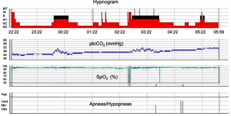
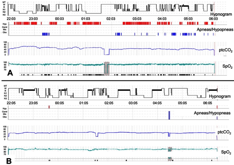
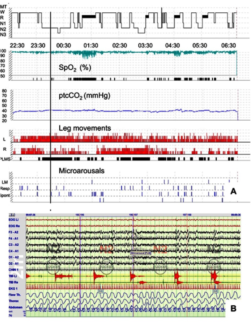
Amyotrophic lateral sclerosis (ALS) is a progressive motor neuron disease inevitably leading to generalized muscle weakness and premature death. Sleep disturbances are extremely common in patients with ALS and substantially add to the burden of disease for both patients and caregivers. Disruption of sleep can be caused by physical symptoms, such as muscle cramps, pain, reduced mobility, spasticity, mucus retention, and restless legs syndrome. In addition, depression and anxiety may lead to significant insomnia. In a small subset of patients, rapid eye movement (REM) sleep behavioral disorder may be present, reflecting neurodegeneration of central nervous system pathways which are involved in REM sleep regulation. With regard to overall prognosis, sleep-disordered breathing (SDB) and nocturnal hypoventilation (NH) are of utmost importance, particularly because NH precedes respiratory failure. Timely mechanical ventilation is one of the most significant therapeutic measures to prolong life span in ALS, and transcutaneous capnometry is superior to pulse oxymetry to detect NH early. In addition, it has been shown that in patients on home ventilatory support, survival time depends on whether normocapnia, normoxia, and elimination of apneic events during sleep can be reliably achieved. Several studies have investigated sleep patterns and clinical determinants of sleep disruption in ALS, but exact prevalence numbers are unknown. Thus, constant awareness for sleep-related symptoms is appropriate. Since no curative treatment can be offered to affected patients, sleep complaints should be thoroughly investigated in order to identify any treatable etiology and improve or stabilize quality of life as much as possible. The use of hypnotics should be confined to palliation during the terminal phase and refractory insomnia in earlier stages of the disease, taking into account that most compounds potentially aggravate SDB.

Keywords: amyotrophic lateral sclerosis; health-related quality of life; non-invasive ventilation; sleep disorders; sleep-disordered breathing.


Conflict of interest statement

Dr Matthias Boentert reports grants from Sanofi-Genzyme GmbH and Loewenstein Medical GmbH, outside the submitted work, and no other conflicts of interest in this work.

Figures

Figure 1
Figure 1
Polysomnography synopsis from a 67-year-old non-ventilated male patient with spinal onset ALS. The figure depicts four tracings representing hypnogram, transcutaneous capnometry, pulse oxymetry, and graphical presentation of apneas or hypopneas (from top to bottom). Whereas capnometry shows hypercapnia which begins during REM and persists during the last third of the night, peripheral oxygen saturation is normal throughout the entire recording. Abbreviations: ALS, amyotrophic lateral sclerosis, REM/R, rapid eye movement sleep, W, wake, N1 - N3, non-REM sleep stages, ptcCO2, transcutaneous carbon dioxide tension, SpO2, peripheral oxygen saturation, Hyp, hypopneas, Cent/Mix/Obs, central/mixed/obstructive apneas.
Figure 2
Figure 2
Polysomnography synopses from a 61-year-old non-ventilated patient with spinal onset ALS and clinically apparent respiratory muscle weakness before and after initiation of non-invasive ventilation (NIV). Both panels depict hypnogram, graphical presentation of apneas and hypopneas, transcutaneous capnometry, and pulse oxymetry (from top to bottom). (A) Sleep fragmentation with reduced sleep efficiency (65.6%), N3 percentage (13.3%), and REM percentage (6.1%). Sleep-disordered breathing is present, comprising severe obstructive sleep apnea (AHI 83.8/h, mean SpO2 88%) and nocturnal hypoventilation (mean ptcCO2 54.0 mmHg, maximum ptcCO2 63.7 mmHg). (B) Rapid improvement of objective sleep outcomes in the very first night of NIV treatment. Normalization of the AHI (0.3/h) and improvement of both CO2 elimination and oxygenation are evident (mean ptcCO2 52.0 mmHg, maximum ptcCO2 55.8 mmHg, mean SpO2 91%). In parallel, sleep architecture shows substantial increase of N3 and REM percentages (25.3% and 26.9%, respectively). Note that sleep-onset REM is present at the beginning of the night. Abbreviations: W, wake, R/REM, rapid eye movement sleep, N1-N3, non-REM sleep stages, ptcCO2, transcutaneous carbon dioxide tension, SpO2, peripheral oxygen saturation.
Figure 3
Figure 3
Periodic limb movements in sleep in a 62-year-old, non-ventilated patient with spinal onset ALS. (A) A polysomnography synopsis including hypnogram, oxygen saturation, transcutaneous ptcCO2, and graphical presentation of both leg movements (LM), and (micro)arousals from sleep (from top to bottom). Whereas capnometry is normal throughout the night, isolated obstructive sleep apnea is present leading to persistent desaturations (AHI 39.8/h). Almost permanent leg movements can be seen in both legs with long-lasting periodicity as reflected by the black bars within the fourth panel from above (PLMS). The PLM index was 117.1/h. However, only few PLM were associated with arousals from sleep, resulting in a PLM arousal index of no more than 5.4/h. (B) A 2-min excerpt from the same polysomnography with the timepoint according to the vertical black line in (A). Sleep stage is N2, and sleep-disordered breathing is absent. Only one of the 8 PLM which can be seen in the electromyography tracings (TIB L and TIB R, respectively) relates with an EEG arousal. Abbreviations: ALS, amyotrophic lateral sclerosis, PLM, periodic limb movemements, ptcCO2, transcutaneous carbon dioxide tension, SpO2, peripheral oxygen saturation, W, wake, R/REM, rapid eye movement sleep, N1-N3, non-REM sleep stages, LM, limb movements, L, left leg, R, right leg, TIB, tibialis anterior muscle, AHI, apnoea hypopnea index.

Similar articles

Cited by

References

    1. Grad LI, Rouleau GA, Ravits J, Cashman NR. Clinical spectrum of amyotrophic lateral sclerosis (ALS). Cold Spring Harb Perspect Med. 2017;7:8. doi:10.1101/cshperspect.a024117 - DOI - PMC - PubMed
    1. Hardiman O, Al-Chalabi A, Chio A, et al. Amyotrophic lateral sclerosis. Nat Rev Dis Primers. 2017;3:17071. doi:10.1038/nrdp.2017.71 - DOI - PubMed
    1. Mizutani T, Aki M, Shiozawa R, et al. Development of ophthalmoplegia in amyotrophic lateral sclerosis during long-term use of respirators. J Neurol Sci. 1990;99(2–3):311–319. - PubMed
    1. Carvalho M, Schwartz MS, Swash M. Involvement of the external anal sphincter in amyotrophic lateral sclerosis. Muscle Nerve. 1995;18(8):848–853. doi:10.1002/mus.880180808 - DOI - PubMed
    1. Hortobagyi T, Cairns NJ. Amyotrophic lateral sclerosis and non-tau frontotemporal lobar degeneration. Handb Clin Neurol. 2017;145:369–381. doi:10.1016/B978-0-12-802395-2.00026-2 - DOI - PubMed